Can Gene Therapy Help Treat Brain Diseases?

Post by D. Chloe Chung

What is gene therapy?

The goal of gene therapy is to alleviate disease symptoms and ultimately cure diseases by correcting abnormal gene expression. This can be done by introducing genetic materials that express exogenous genes or suppress the expression level of endogenous genes in an effort to modify gene expression levels. Gene therapy can be also designed to directly edit gene mutations present in patients. Over the past years, development of novel gene-editing tools has resulted in improved efficacy of gene delivery to the brain. With this exciting technical advancement, gene therapies have recently gained more attention as a potential therapeutic strategy for neurodevelopmental and neurodegenerative diseases, especially for those caused by genetic mutations that disrupt the body’s usual patterns of gene expression.

Different strategies behind gene therapy

There are several ways to design gene therapy to correct aberrant gene expression in neurological diseases. One of them is to express exogenous proteins that can restore the function of faulty endogenous proteins. For this purpose, the adeno-associated virus (AAV) is a preferred viral vector because of its relative safety as well as its long-term gene expression which reduces the need for repeated administration. After being introduced into cells, AAVs can express genes that they carry by using the transcriptional and translational machinery in the host cells. In this way, AAVs produce proteins that can make up for the loss of gene function. The virus engineering field continues to refine structures of AAVs to increase their safety as well as effectiveness in delivery to the central nervous system.

DNA-editing is also an appealing approach for gene therapy as it can directly fix disease-causing mutations and modify gene expression level. The newest advancement in DNA-editing tools involves the synthetic CRISPR system (an abbreviation for ‘clustered regularly interspaced short palindromic repeats’) in which a customized guide RNA can bring a DNA-cutting enzyme (e.g., Cas9) to the specific site within the gene of interest. Once the enzyme cuts out a few base pairs from the gene, the gene sequence will eventually shift and create a premature stop codon). The cellular mechanism takes this stop codon as a sign to degrade messenger RNAs transcribed by this gene, ultimately silencing the expression of the target gene. This genetic tool has great potential to treat neurological diseases as it can be used to inactivate or activate genes of interest, or to edit precise bases within the gene and correct pathogenic gene mutations.

In addition to DNA-editing tools, the RNA-based therapy utilizing antisense oligonucleotides (ASOs) has gained much attention for its potential efficacy. ASOs are short DNA or RNA fragments that can bind to messenger RNAs based on the complementary sequence, subsequently changing the RNA expression level. As such, ASOs can be synthesized to target messenger RNAs transcribed from the disease gene of interest in hopes to regulate the protein level that could play crucial roles in neurological diseases.

How is gene therapy treatment for neurological diseases going?

In 2017, a groundbreaking study published in The New England Journal of Medicine reported the successful usage of gene therapy in young children with spinal muscular atrophy (SMA) type 1, a devastating neuromuscular disease characterized by motor neuron degeneration and progressive muscle loss. In SMA, a defective gene SMN1 reduces the amount of functional SMN1 protein, so researchers treated SMA patients with a one-time blood infusion of AAV that can express the SMN1 gene and restore protein expression level. Excitingly, most of the patients who received this gene therapy showed drastic improvement in their survival and motor functions that lasted through the 2-year follow-up assessment. Some of the patients were even able to walk with no assistance, which is a striking development considering that many SMA patients need wheelchair assistance and die at a very young age.

As of August 2021, a medication designed to increase the level of SMN protein is the only disease-modifying gene therapy approved by the US Food and Drug Administration (FDA) to treat neurological symptoms. Yet, in addition to this medication, numerous preclinical and clinical studies are actively investigating the safety and efficacy of gene therapy for a wider range of neurological diseases. For example, ASOs targeting the gene that makes tau, a protein that becomes abnormally aggregated in Alzheimer’s disease, have been tested in mouse models. After demonstrating their ability to reduce tau pathology and subsequently rescue behavioral deficits in a mouse model, the ASO-mediated tau-targeting gene therapy is being tested in a clinical trial for Alzheimer’s disease patients.

Similarly, ASO-based gene therapy has been investigated for its potential benefits in patients of Huntington’s disease, which is caused by an abnormal trinucleotide expansion in the huntingtin gene. The idea behind this therapy is to use ASOs either to globally target the total huntingtin gene level or to specifically reduce the function of the mutant allele. Based on promising results from preclinical studies, several ASOs entered clinical trials to evaluate their safety and efficacy for Huntington’s disease patients. Unfortunately, however, a couple of these clinical trials have been recently discontinued due to lack of evidence of anticipated benefits for patients.

What are the challenges and what is the hope for the future?

Perhaps the biggest challenge in utilizing gene therapy to treat neurological diseases is safety. For example, when genetic materials are being delivered at a high dosage, they can cause toxicity in patients. Also, undesirable immune responses can occur upon introduction of genetic materials. As these scenarios can lead to fatal consequences, safety is always the first aspect to consider and monitor when designing gene therapy and testing it in clinical trials. Moreover, off-target effects – unwanted changes in genes that were not the target of gene therapy – are also a potential concern. This can happen when ASOs or gene-editing tools target alternative genes based on partially complementary sequences. How to avoid off-target effects can be a major challenge when designing the reagents.

Despite these many challenges, biomedical techniques that improve our ability to utilize gene therapy to effectively treat neurological diseases continue to advance. For instance, delivery of AAVs into the central nervous system used to be highly challenging in the past, since it is not feasible to directly inject AAVs into the brain. At the same time, the blood-brain barrier firmly isolates the brain from the periphery, making it difficult for locally delivered reagents (e.g., injection into blood vessels) to bypass this physical barrier and reach the brain. Yet, characterization of various AAVs and persistent efforts in virus engineering allowed for development of a specific AAV that can be successfully and non-invasively (e.g., without surgeries) delivered to the central nervous system. This type of AAV was also used in designing the effective gene therapy for SMA patients as described above, demonstrating that technological advancement can lead to breakthrough therapeutic strategies. With rapidly evolving gene-editing tools, it's an exciting time for the development of gene therapy for neurological diseases.

References

Sun & Roy. Gene-based therapies for neurodegenerative diseases. Nature Neuroscience (2020). Access the original scientific publication here.

Martier & Konstantinova. Gene therapy for neurodegenerative diseases: slowing down the ticking clock. Frontiers in Neuroscience (2020). Access the original scientific publication here.

Bennett et al. Antisense oligonucleotide therapies for neurodegenerative diseases. Annual Review of Neuroscience (2020). Access the original scientific publication here.

Mendell et al. Single-dose gene-replacement therapy for spinal muscular atrophy. The New England Journal of Medicine (2017). Access the original scientific publication here.

DeVos et al. Tau reduction prevents neuronal loss and reverses pathological tau deposition and seeding in mice with tauopathy. Science Translational Medicine (2017). Access the original scientific publication here.

Kordasiewicz et al. Sustained therapeutic reversal of Huntington’s disease by transient repression of huntingtin synthesis. Neuron (2013). Access the original scientific publication here.

Southwell et al. In vivo evaluation of candidate allele-specific mutant huntingtin gene silencing antisense oligonucleotides. Molecular Therapy (2014). Access the original scientific publication here.

Kwon. Failure of genetic therapies for Huntington’s devastates community. Nature News (2021). Access the news article here.

Hudry & Vandenberghe. Therapeutic AAV Gene Transfer to the Nervous System: A Clinical Reality. Neuron (2019). Access the news article here.

Doudna. The promise and challenge of therapeutic genome editing. Nature (2020).Access the news article here.

The Relationship Between the Menstrual Cycle and Sleep

Post by Shireen Parimoo

What is the menstrual cycle?

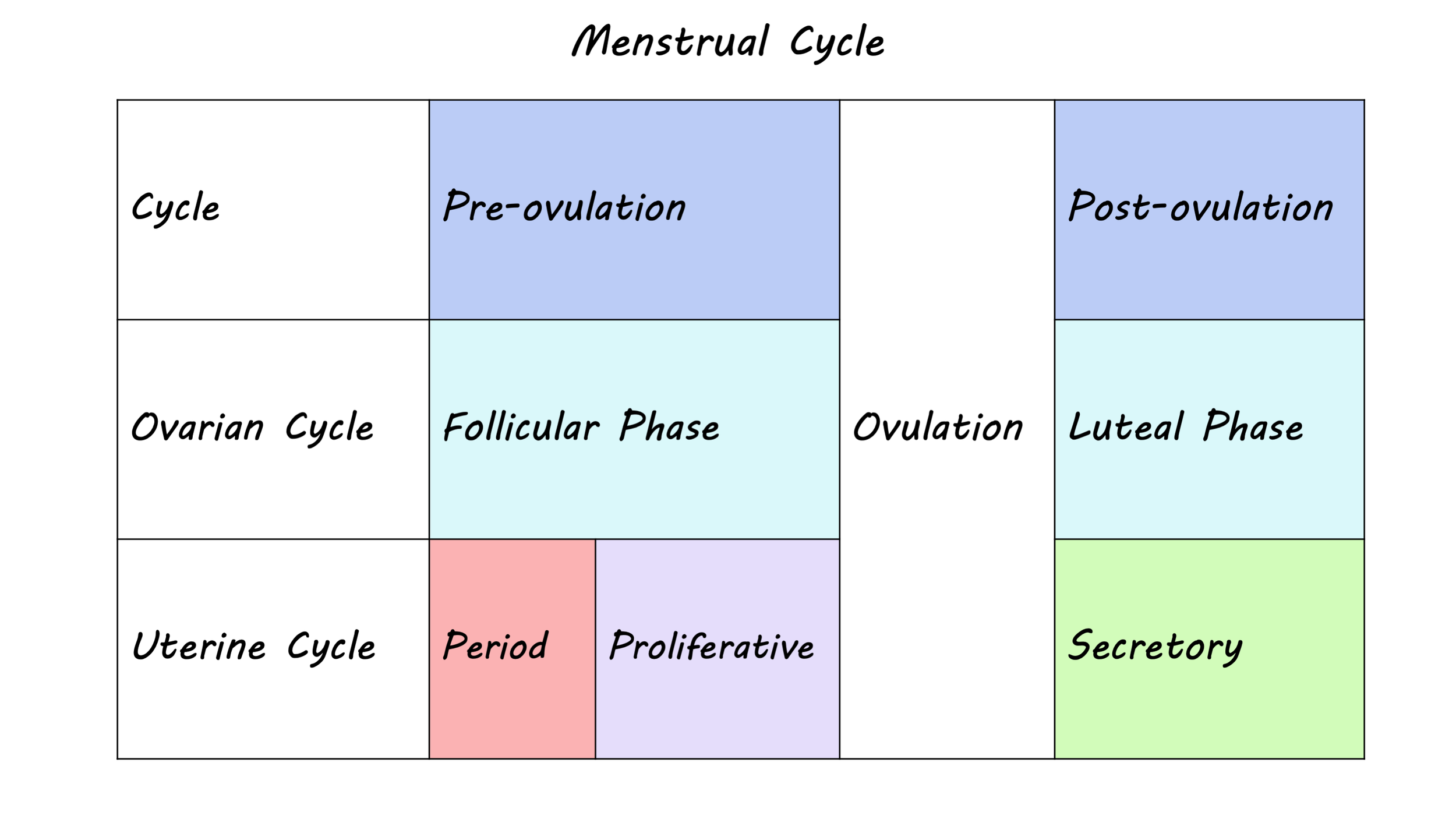
The menstrual cycle describes the fluctuation of ovarian hormones that typically occurs over a 28-day period, but can range anywhere from 21 to 38 days. The cycle is divided into two distinct phases: the follicular phase and the luteal phase. The follicular phase begins on the first day of menses - more commonly known as the period - which lasts between three to eight days. This phase ends with ovulation, when an egg is released from the ovary into the uterus, followed by the luteal phase, which lasts until the next menses.

In each phase of the menstrual cycle, hormonal changes result from an interaction between the brain and the reproductive system. In the follicular phase, follicle stimulating hormone released from the pituitary gland in the brain prepares the ovaries for ovulation and triggers the release of estrogens, which prepares the uterus for ovulation. Toward the end of the follicular phase, high levels of estrogens act on the brain to facilitate the release of luteinizing hormone, which in turn triggers ovulation. Once the egg is released, there is an increase in progesterone and estrogen but if pregnancy does not occur levels decline, leading to the next menses.

Cycle_Jul20.png

How does it affect sleep?

Sleep promotes physical and mental recovery, maintenance, and repair within the brain, and facilitates learning and memory. Thus, it is important to maintain a consistent sleep schedule and get good quality sleep, since sleep disturbances can not only interfere with day-to-day functioning and general well-being but can also disrupt cognitive performance and increase the risk of disease and dementia. Good sleep hygiene, such as keeping a consistent bedtime/nighttime routine, getting enough sunlight during the day, exercising regularly, and avoiding nicotine, alcohol, and stimulants in the evening can help. In general, women self-report more frequent sleep problems, increased daytime sleepiness, and poorer quality of sleep compared to men, yet objective measures like sleep duration suggest that women get more quality sleep than men.

Ovarian hormones partially contribute to these conflicting sex differences in objective and subjective measures of sleep quality. Across the female lifespan, the most pronounced hormonal changes take place during puberty, menses, pregnancy, and menopause, which coincide with sleep disturbances. During reproductive years, women experience more subtle fluctuations in their quality of sleep over the course of the menstrual cycle. Sleep disturbances are primarily observed during the luteal phase, partly due to elevated levels of estrogens and progesterone. For example, women report higher daytime sleepiness and more awakenings at night during the luteal compared to the follicular phase. Core body temperature at night is also elevated during the luteal phase, which is related to both hypersomnia (excessive sleep) and insomnia (inability to sleep). However, some objective measures of sleep quality such as total time spent sleeping and sleep efficiency are not consistently affected by the menstrual phase.

How is the brain involved?

Sleep consists of recurring cycles that usually last about 90 minutes, with one night of sleep involving between three to five sleep cycles. Each sleep cycle includes rapid eye movement (REM) sleep, when dreaming occurs, and non-REM sleep, which involves light and deep sleep. Polysomnography studies show that the duration of REM sleep is lower during the luteal phase than the follicular phase, whereas the duration of non-REM sleep becomes longer.

Ovarian hormones are increasingly being recognized for their relevance in non-reproductive functions through their impact on the brain. For example, there is evidence of better memory after a nap for women in the luteal phase compared to those in the follicular phase of their menstrual cycle. Estrogens and progesterone, which are elevated during the luteal phase, act on receptors in the hippocampus and the frontal cortex, regions that are involved in learning, memory, and decision-making. Moreover, these hormones have also been shown to have neuroprotective effects on the brain’s structure across the lifespan.

Slow-wave sleep (deep sleep) and sleep spindles are prominent features of non-REM sleep, which is when much of the sleep-related recovery and memory consolidation takes place. Sleep spindles refer to bursts of oscillatory activity (11-16 Hz) that typically occur during stage 2 of non-REM sleep. Notably, sleep spindles occur more frequently during the luteal phase of the menstrual cycle and are associated with memory performance. Higher levels of progesterone are thought to modulate spindle activity by acting on GABA receptors in the brain. Conversely, slow-wave sleep is characterized by low frequency oscillatory activity (0.5-3 Hz) and is reduced during the luteal phase. However, it is currently unclear how brain activity during different sleep stages is linked to specific phases of the menstrual cycle. More research is needed to better understand the complex and interdependent relationship between the female reproductive system and brain in relation to sleep and cognition. 

References

Alonso et al. Sex and menstrual phase influences on sleep and memory. Current Sleep Medicine Reports (2021).

Baker & Driver. Self-reported sleep across the menstrual cycle in young, healthy women. Journal of Psychosomatic Research (2004).

Bixler et al. Women sleep objectively better than men and the sleep of young women is more resilient to external stressors: the effects of age and menopause. Journal of Sleep Research (2009).

Brann et al. Neurotrophic and neuroprotective actions of estrogen: basic mechanisms and clinical implications. Steroids (2007).

Brinton et al. Progesterone receptors: form and function in the brain. Frontiers in Neuroendocrinology (2008).

Brown & Gervais. Role of ovarian hormones in the modulation of sleep in females across the adult lifespan. Endocrinology (2020).

Dorsey et al. Neurobiological and hormonal mechanisms regulating women’s sleep. Frontiers in Neuroscience (2021).

Driver et al. The menstrual cycle effects on sleep. Sleep Medicine Clinics (2008).

Genzel et al. Sex and modulatory menstrual cycle effects on sleep related memory consolidation. Psychoneuroendocrinology (2012).

Gervais et al. Ovarian hormones, sleep and cognition across the adult female lifespan: an integrated perspective. Frontiers in Neuroendocrinology (2017).

Johnson et al. Epidemiology of DSM-IV insomnia in adolescence: lifetime prevalence, chronicity, and an emergent gender difference. Pediatrics (2006).

Mong & Cusmano. Sex differences in sleep: impact of biological sex and sex steroids. Philosophical Transactions of the Royal Society B (2016).

Plante & Goldstein. Medoxyprogesterone acetate is associated with increased sleep spindles during non-rapid eye movement sleep in women referred for polysomnography. Psychoneuroendocrinology (2013).

Romans et al. Sleep quality and the menstrual cycle. Sleep Medicine (2015).

Schumacher et al. Progesterone: therapeutic opportunities for neuroprotection and myelin repair. Pharmacology and Therapeutics (2007).

De Zambotti et al. Menstrual cycle-related variation in physiological sleep in women in the early menopausal transition. Journal of Clinical Endocrinology and Metabolism (2015).

Does Sleep “Clean” Our Brains?

Post by Lani Cupo

Why do we spend so much of our life asleep?

An Irish proverb states: “A good laugh and a long sleep are the best cures in the doctor’s book.” We spend almost one-third of our lives asleep, but what makes sleep restorative, and why do we need so much of it? Sleep has many benefits like facilitating memory consolidation and emotion regulation, but today we focus on the role sleep plays in clearing the brain of neurotoxins that accumulate during waking hours. During daily functioning, the brain accumulates proteins such as β-amyloid (Aβ), α-synuclein, and tau over the course of waking hours. Accumulation of these proteins over the lifespan may contribute to brain pathology, so it is important that concentrations of these proteins are regulated. Recent evidence suggests that sleep may in part fill this role, helping to protect the brain by clearing the excess of these proteins.

What do we know?

Brain tissue is composed of three main components: neural cells, vasculature, and the interstitial system (ISS) referring to the space between cells and blood vessels. Most recent research on the brain focuses on cells such as neurons and glia, however, the ISS forms the microenvironment of the brain. It occupies 15-20% of total brain volume and plays a pivotal role in healthy brain functioning. Evidence from mice suggests that during sleep, interstitial volume can increase up to 60% allowing for increased flow between interstitial fluid and cerebrospinal fluid (CSF), the fluid in which the brain is floating. This might facilitate the improved clearance of toxins from brain tissue. While the mechanism allowing for the change in volume is still unknown, one hypothesis is that support cells known as astrocytes shrink during sleep, resulting in the observed volumetric changes.

To examine whether sleep facilitates the clearance of metabolites via CSF, one study in humans injected individuals with a CSF tracer they could image with magnetic resonance imaging (MRI) and investigated the impact of acute (one night) sleep deprivation on tracer clearance as a proxy for metabolite clearance. Following a night of sleep deprivation, tracer clearance was reduced, suggesting less effective clearance of neurotoxins. This finding is significant not only because it presents some of the first live human evidence, but also because the authors were able to assess clearance in deep structures within the brain.

During wakefulness, ISS contraction increases tissue resistance, reducing the influx of CSF. This potentially alters not only the clearance of excess neurotransmitters but also aggregates of proteins in the brain. Circadian rhythms, which help regulate sleep cycles, may also impact clearance by altering the permeability of the blood-brain barrier, the interface between circulating blood and the central nervous system. During sleep, this barrier becomes more porous, further impacting the clearance of proteins. Examining the clearance of the Aβ protein, one study in mice found the protein was cleared twice as fast during sleep as compared to wakefulness. This holds important implications for neurodegenerative disorders, as the accumulation of Aβ plaques is a hallmark of Alzheimer’s Disease (AD) pathology.

What are the implications for Alzheimer’s Disease?

Sleep is an important factor in the emergence of neurodegenerative disorders, such as AD and Parkinson’s Disease. When there is an imbalance between Aβ production and clearance in the brain, the protein can stick together causing aggregates, known as plaques, to form. Excess tau protein can also get stuck together forming “tangles”. The formation of Aβ plaques and tau tangles contribute to the loss of neurons and their connections. Similar to human studies, rodent models show that sleep deprivation elevates concentrations of Aβ, with concentrations increasing consistently over prolonged sleep deprivation. While an increased risk for AD has been associated with a shorter duration of sleep, the causal link between sleep deprivation and heightened risk for AD remains to be determined. It also remains unclear whether the mechanistic link between sleep disturbances and AD involves neurotoxin clearance.

The specific mechanism of toxin clearance from the brain is still unknown, although preliminary research implicates a specific water channel known as aquaporin-4 in the removal of interstitial waste. Recent studies implicate a brain region known as the locus coeruleus (LC) in the regulation of sleep - signaling from the LC is associated with states of wakefulness. This region displays volumetric abnormalities in AD, suggesting that it may be related to the pathophysiology of the disease.

What is the takeaway message?

During the time we sleep our brain tissues undergo changes that facilitate more efficient cleansing of the toxins and waste that naturally accumulate in our brains over the course of the day. This mechanism could underlie the observed association between neurological disorders like AD and sleep disturbance, however, it remains unclear whether sleep deprivations exacerbate AD pathology or if AD pathology exacerbates sleep disruption. Of course, if you don’t get enough sleep it does not mean that you will develop a neurological disorder, however, the research strongly suggests that sleep is a critical factor in brain health. Overall, the benefits of sleep are many-fold, and we are still learning exactly how sleep supports and protects our brain.


References

Albrecht, et al. Circadian Clocks and Sleep: Impact of Rhythmic Metabolism and Waste Clearance on the Brain. Trends in Neurosciences. (2018). Access the original scientific publication here.

Eide, et al. Sleep Deprivation Impairs Molecular Clearance from the Human Brain. Brain: Journal of Neurology. (2021). Access the original scientific publication here.

Goldstein & Walker. The role of sleep in emotional brain function. Annu Rev Clin Psychol. (2014). Access the original scientific publication here.

Huang, et al. Sleep, Major Depressive Disorder and Alzheimer’s Disease: A Mendelian Randomisation Study. Neurology. (2020). Access the original scientific publication here.

Lei, et al. The Brain Interstitial System: Anatomy, Modeling, in Vivo Measurement, and Applications. Progress in Neurobiology (2017). Access the original scientific publication here.

Mendelsohn & Larrick. Sleep Facilitates Clearance of Metabolites from the Brain: Glymphatic Function in Aging and Neurodegenerative Diseases. Rejuvenation Research (2013). Access the original scientific publication here.

Xie, et al. Sleep Drives Metabolite Clearance from the Adult Brain. Science (2013).Access the original scientific publication here.