Eating Highly Processed Foods is Associated with Stroke and Cognitive Impairment

Post by Shahin Khodaei

The takeaway

Eating more heavily processed foods is associated with an increased risk of cognitive decline and stroke. On the flip side, eating more unprocessed or minimally processed foods is associated with a decreased risk of cognitive decline and stroke. 

What's the science?

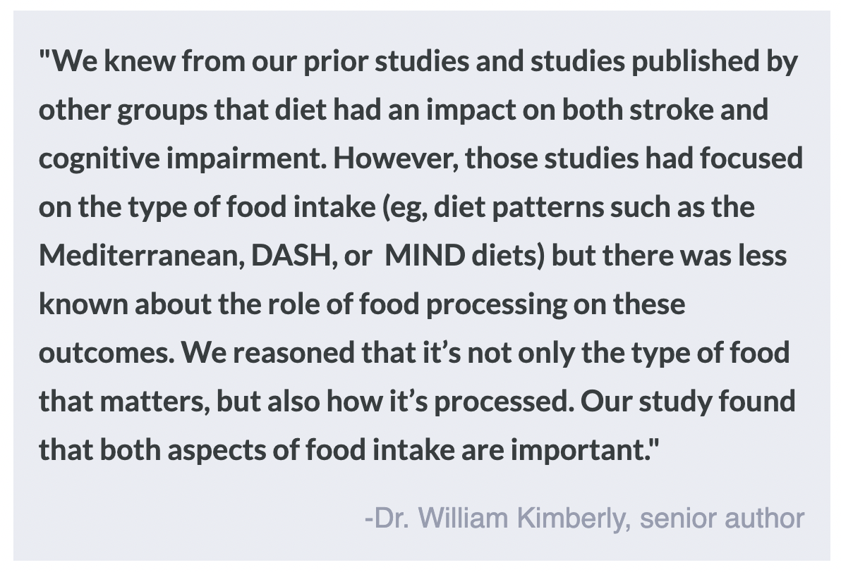
Diet is known to affect the brain - for example, following a more Mediterranean diet is associated with a reduced risk of stroke and lower cognitive decline. Recent research also indicates that eating more ultra-processed foods (e.g. carbonated drinks, flavoured yogurt, instant foods, packaged bread, chicken nuggets, etc.) is associated with a higher risk of stroke and faster cognitive decline. This week in Neurology, Bhave and colleagues published a study that adds to the growing literature on diet and brain health outcomes, investigating the role of food processing compared to following specific diets such as the Mediterranean diet. 

How did they do it?

The authors followed a cohort of over 30,000 non-Hispanic Black and White adults aged 45 and above in the United States, who entered the study between 2003 and 2007. After enrolling in the study, participants were assessed for clinical information, including a history of stroke or cognitive impairments, and demographic and lifestyle information. During the baseline assessment, participants answered a questionnaire about their food intake, which was then analyzed in two ways: 1) The food and drink items were categorized into four groups based on the level of processing, and daily intake for each category (in grams) was divided by total intake to get a proportion. 2) the questionnaires were scored based on how much they adhered to three healthy dietary patterns – Mediterranean, Dietary Approaches to Stop Hypertension (DASH), and Mediterranean-DASH Intervention for Neurodegenerative Delay (MIND).

After this baseline assessment, participants were followed up routinely to assess whether they had experienced a stroke, and to assess their cognitive performance in standardized tests. The authors then built statistical models to investigate the associations between food and drink intake and the incidence of stroke and cognitive impairment. 

What did they find?

The study found that eating more ultra-processed foods was associated with an increased risk of stroke, particularly in Black participants. On the other hand, eating more unprocessed/minimally processed foods or more strongly following a healthy diet was associated with a decreased risk of stroke. The results were similar when the authors looked at cognitive impairment – more ultra-processed foods were associated with greater cognitive decline, while less processed foods and healthier diets were associated with less decline.

The authors also asked a follow-up question: does the level of food processing matter if participants are following a healthier dietary pattern such as DASH or MIND? The answer was yes: even when participants adhered to a healthy diet, eating more ultra-processed foods was associated with some negative brain health outcomes, and eating more unprocessed foods was associated with better outcomes. This finding suggests the level of processing in the diet alone, is important for brain health, independently of other dietary patterns.

What's the impact?

This study highlights the important role that food processing plays in brain health. As always, it is important to note that these findings do not necessarily mean that eating more processed foods directly causes stroke and cognitive impairment (i.e. correlation is not causation). However, this study contributes to a growing literature that suggests a healthy diet including unprocessed food is important in maintaining brain health. 

Neuron to Neuron Information Transfer is Critical for Emotion Recognition and Social Cognition

Post by Soumilee Chaudhuri

The takeaway

Information transfer from the medial prefrontal cortex (mPFC) to the retrosplenial cortex (RSC) of the brain is crucial for emotion recognition - the ability to recognize and respond to the emotional states of others. This study found that inhibiting the mPFC-to-RSC brain pathway in mice affects their ability to recognize emotional states like stress and relief in their peers, shedding light on the neural mechanisms behind social cognition.

What's the science?

Emotion recognition is essential for appropriate social interactions, enabling individuals to respond to the emotional states of others. Recent research has revealed that recognizing emotions in others involves complex communication between different parts of the brain, but understanding the exact brain pathways involved has been challenging. This study in Nature Neuroscience by Dautan et. al., delves into the mPFC-to-RSC pathway, focusing specifically on the role of somatostatin (SOM) neurons that project from the mPFC to the RSC. SOM neurons are known for their inhibitory role in the brain - they produce Gamma-aminobutyric acid (GABA), an inhibitory neurotransmitter. It is also known that these SOM neurons help filter and process socially derived information, enabling accurate emotion recognition. In this study, researchers used optogenetics to manipulate the mPFC-to-RSC SOM neurons and observe their impact on the behavior of the mice.

How did they do it?

In this study, researchers utilized a combination of behavioral tests, optogenetics, and calcium imaging to investigate how information transfer between the mPFC and RSC affects emotion recognition in mice. The focus was on the interaction between specific neuron types, including pyramidal neurons in the RSC and SOM neurons in the mPFC. They specifically targeted genetically modified SOM neurons that expressed light-sensitive proteins. Light was used to either activate or inhibit these neurons and behavioral tests in mice assessed their emotions during these periods of activation or inhibition. Behavioral tests in mice included a series of assessments wherein mice had to recognize emotional states (stress or relief) in other mice. By stimulating or inhibiting the SOM neurons in the mPFC, researchers could observe changes in the mice's ability to recognize these emotional cues and record their responses.

What did they find?

The researchers found that inhibiting the mPFC-to-RSC pathway impaired the mice's ability to recognize emotions in their peers while stimulating it enhanced their emotional recognition capabilities. The results indicated that SOM neurons in the mPFC regulate the activity of RSC pyramidal neurons, influencing how mice process and respond to social and emotional stimuli. When the mPFC-to-RSC SOM neurons were inhibited, the activity of RSC pyramidal neurons increased, indicating that the SOM neurons help regulate the signal-to-noise ratio within the RSC. This regulation of mPFC SOM neurons to RSC pyramidal neurons was vital for processing and interpreting social and emotional stimuli accurately and it was shown that about 10% of mPFC SOM neurons project to RSC and thus modulate the activity of RSC pyramidal neurons. Interestingly, these results in mice were similar to what other scientists have observed in recent functional magnetic resonance imaging(fMRI) studies in humans.

What's the impact?

Understanding the mPFC-to-RSC pathway's role in emotion recognition has significant implications for studying social cognitive disorders like autism and schizophrenia, where emotion recognition is often impaired. This research offers avenues for therapeutic strategies targeting specific brain pathways to improve social functioning. Additionally, it provides a deeper understanding of the neural mechanisms underlying social behavior and emotional processing, which could inform future studies in both animals and humans. 

Access the original scientific publication here.

Modeling the Dose-Dependent Effects of Ketamine

Post by Lani Cupo

The takeaway

Ketamine produces sedation and disassociation at low doses and anesthesia at high doses accompanied by specific patterns of brain activity characteristic of each state. Disinhibition of neural circuits leading to a global increase in excitation may underlie both low and high-dose states.

What's the science?

The dose-dependent effects of ketamine are well known, with low doses producing psychoactive effects and high doses producing anesthesia. Likewise, it is known that ketamine administration produces patterns of brain activity consistent with gamma oscillations (associated with cognitive function) at low doses, but these are interrupted by slow-delta oscillations (associated with deep sleep) at higher doses. Nevertheless, it’s still an open question how cellular processes relate to the emergence of these patterns of brain activity. This week in PNAS, Adam and colleagues present a biophysical model to simulate cellular changes and observe the effect on brain oscillatory behavior, finding that interactions between inhibitory and excitatory neurotransmitters play a role in the distinctive patterns of brain oscillations observed following ketamine exposure.

How did they do it?

First, the authors acquired electroencephalogram (EEG) data from a human volunteer and a nonhuman primate who were administered ketamine at doses sufficiently high to induce anesthesia. Then, they created a biophysical model (a simulation of biological processes) representing interactions between excitatory pyramidal neurons and inhibitory interneurons. The model focused on the activity of NMDA receptors (a major receptor of interest for ketamine), allowing NMDA receptors to change state (“open” ones can become “closed”) based on other activity in the system. Specifically, in biology, ketamine is known to block the excitatory NMDA receptors, which is interesting given the fact that low levels of ketamine create an excitatory state. It is thought that this is because ketamine blocks inhibitory neurons from firing, leading to an overall excitatory state. The authors tested this hypothesis in their biophysical model. Then, they examined what changes in the activity of neurons could explain gamma oscillations seen following ketamine exposure. Next, they examine why slow-wave delta oscillations emerge when ketamine is “increased” in the model. 

What did they find?

First, the authors found characteristic patterns of EEG activity: at low levels of ketamine, gamma oscillations, representing cortical activity, were evident, whereas at higher levels of ketamine exposure, gamma oscillations were interrupted by delta waves, characteristic of sleep.

Then, using the biophysical model, the authors found evidence for the cellular mechanisms contributing to the gamma oscillations. They found evidence that ketamine blocked NMDA receptors on inhibitory interneurons, contributing to an overall excitatory state. Specifically, some neurons have a subthreshold excitatory state, meaning at baseline they are close to firing, but not quite over the threshold that makes them fire. Blocking these neurons’ NMDA receptors with ketamine can shut them down. When these neurons release inhibitory neurotransmitters, shutting them down leads to a downstream increase in excitatory neurotransmitter release, or a global increase in excitation referred to as disinhibition, because the excitatory neurons are no longer inhibited.

With their biophysical model, the authors next observed that this global excitation gave rise to gamma patterns of brain activity. This behavior is dependent on inhibitory GABA-ergic neurons, some of which are not blocked by ketamine, which can contribute to individual neurons firing at a gamma timescale. These individual neurons are synchronized across the brain, giving rise to global gamma wave activity.

In their model, the authors also find that higher doses of ketamine can induce “down-states” associated with slow-wave delta oscillations. Neurons with background excitatory states shut down under increased ketamine administration while other neurons have a reduced timescale of firing, contributing to the slower delta waves.

What's the impact?

The authors demonstrate that ketamine can produce characteristic brain waves in a biophysical model by blocking NMDA receptors. Their findings increase our understanding of the cellular mechanisms contributing to global brain activity.

 Access the original scientific publication here.