The Role of Frontal Theta-Band Activity in Regulating Risk-Taking Behavior

Post by Kulpreet Cheema

The takeaway

Previous research suggests that frontal cortex theta-band activity - a rhythm of neural activity between 4-8 Hz - plays a significant role in modulating decision-making involving risk. Theta-band activity in the left dorsolateral prefrontal cortex (DLPFC) specifically, is associated with increased risk-taking behavior. 

What's the science?

We face risky choices constantly in everyday life, from small decisions like whether to take an umbrella out with us based on the weather forecast, to larger decisions like deciding how to invest our money in the stock market. Electroencephalography (EEG) studies have shown that frontal theta-band activity is associated with cognitive control, response inhibition, reward anticipation, and conflict detection processes that are essential in risk-taking behavior. The dorsolateral prefrontal cortex (DLPFC) and medial prefrontal cortex (MPFC) are two crucial frontal brain regions involved in decision-making, particularly in a risky context. This week in NeuroImage, Dantas and colleagues aimed to understand the role of frontal cortex theta-band activity in regulating risk-taking behavior. 

How did they do it?

Thirty-nine participants performed the Maastricht Gambling Task (MGT) while their right or left DLPFC were stimulated with transcranial alternating current stimulation (tACS). tACS is a unique form of brain stimulation used to modulate brain activity and in some cases induce neural plasticity. The MGT was carefully selected to assess risk-taking behavior, controlling for the effects of other variables like loss aversion and memory and learning effects. In each trial, participants had to guess the color of a box hiding a token, with varying probabilities of success and corresponding payoffs. Two tACS stimulation intensities (1.5 mA and 3 mA) were applied to assess the impact of intensity on risk-taking. The researchers analyzed behavioral data, including risk, value, and response time, as well as EEG data, to understand how brain stimulation affected risk-taking behavior. 

What did they find?

The results showed that stimulating the left DLPFC with theta-band tACS led to increased risk-taking behavior. However, when the right DLPFC was stimulated, no significant changes in risk-taking were observed. Participants' ‘value’, which reflects their attraction to larger payoffs or rewards in a given trial of the gambling task, increased significantly during left DLPFC stimulation, especially with higher intensity. In contrast, right DLPFC stimulation, particularly at higher intensity, led to a significant reduction in value. EEG data revealed increased theta power after sham stimulation and an overall increase in frontal theta power after left DLPFC tACS. However, no significant EEG aftereffects were observed following right DLPFC tACS. 

What's the impact?

This study sheds light on the differential role of right and left frontal theta-band activity in risk-taking behavior. Researchers found that stimulating specific brain regions can influence decision-making under risk, with left DLPFC stimulation increasing risk-taking, and right DLPFC stimulation reducing it, particularly at higher intensities. Understanding these relationships has potential applications in clinical settings, as these findings could inform intervention in cases involving abnormal risk-taking behavior. The research also highlights the importance of tACS stimulation intensity, indicating that higher intensities may be necessary to induce consistent behavioral responses. 

Access the original scientific publication here.

A Less Invasive Form of Deep Brain Stimulation Enhances Memory Function in Healthy Adults

Post by Christopher Chen 

The takeaway

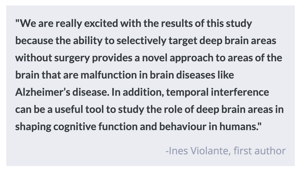
Deep brain stimulation (DBS) can be used to treat neurological and psychiatric disorders, but the technique can be invasive and cause unwanted side effects. Scientists have developed a non-invasive method of DBS called temporal interference (TI) and demonstrated its effectiveness in humans by focusing on the hippocampus, where TI helped improve memory accuracy in healthy subjects.  

What's the science?

Since its FDA approval in 1997 in the treatment of essential tremor, deep brain stimulation (DBS) – which relies on direct electrical stimulation of specific brain regions – has since been approved and used by clinicians to treat patients with neurological conditions such as Parkinson’s disease, epilepsy, and most recently, Alzheimer’s disease. However, DBS is quite invasive, with its use requiring patients to undergo brain surgery which can lead to additional complications. As such, researchers have sought safer, non-invasive forms of DBS such as transcranial magnetic stimulation and transcranial electrical stimulation (tES) to treat brain-related disorders. Unfortunately, these techniques can also elicit harmful side effects through unintended activation of brain areas close to but not part of the target stimulation region. 

Recently, scientists have developed a method called temporal interference (TI) stimulation, which uses multiple electric fields delivered at different frequencies to target specific brain regions. By changing the distribution of current intensities between different scalp electrodes, researchers can also direct, or steer, TI to different areas. This technique offers a way to stimulate deep brain structures without the risks of surgery, while also providing a level of precision not found in other tES techniques.

Recently in Nature Neuroscience, Violante and colleagues described how scientists used TI to perform non-invasive, targeted stimulation of the hippocampus in humans under several different conditions. Data also demonstrated that TI-mediated stimulation of the hippocampus augmented memory function in healthy human subjects, highlighting the potential therapeutic benefits of TI in the near future. 

How did they do it?

Considering one of the main drawbacks of other forms of neuromodulation is non-specificity, the researchers wanted to see if they could apply TI to an exclusive region of the hippocampus without disturbing the overlying cortex as well as reliably steer TI to other parts of the hippocampus. To test this, the researchers relied on an anatomical model of the human brain that could measure the effects of electrical field stimulation. They measured electrical field differences between the hippocampus and overlying cortex as well as the ability of the signal to be steered to different parts of the hippocampus. Following application in computer models, this experimental paradigm was applied to human cadavers.   

The researchers used neuroimaging in live human subjects to assess the physiological impact of TI stimulation on the hippocampus. To do so, they applied TI to 20 healthy participants while monitoring their brain activity via functional magnetic resonance imaging (fMRI). Specifically, they transiently applied TI stimulation on the hippocampus during a face-name paired associative task known to measure the encoding of episodic memory and measured the evoked hippocampal blood-oxygen-level-dependent (BOLD) signal in different hippocampal regions. 

A subsequent behavioral experiment involved a new cohort of 21 participants undergoing a similar face-name associative task paired with fMRI, albeit with a longer TI stimulation duration. This experiment was designed to probe the behavioral consequences of TI stimulation on memory, and stimulation was applied during the encoding, maintenance, and recall stages. 

What did they find?

Electric field modeling using computer simulations as well as measurements in a human cadaver validated the ability to localize TI stimulation to the human hippocampus with minimal exposure to the overlying cortex. The electric fields generated by TI also had amplitudes consistent with previous computational studies and were within the range to synchronize neural spiking activity in the desired frequency range.

Results from the neuroimaging experiments in live human subjects showed that TI stimulation, when transiently applied during the encoding of episodic memory, reduced the BOLD signal. Importantly, this reduction was specific to the hippocampus, with no significant effect on the overlying cortex. The strongest reduction occurred when TI stimulation was targeted to the anterior hippocampus, which plays a central role in memory encoding. These data collectively show that TI can be successfully localized to a specific region of the hippocampus known to be involved in memory formation.  

In the behavioral experiments involving longer stimulation durations and multiple components of memory processing, data showed a small but significant improvement in memory accuracy in participants when TI was applied. However, memories formed during TI stimulation were forgotten at a rate similar to those during sham stimulation. Overall, data suggests that TI-mediated augmentation of memory may be specific to memory accuracy. 

What's the impact?

This study is the first to characterize the functionality of TI in live human subjects and show that TI can be localized to the anterior hippocampus. As for TI’s functional significance, this study suggests that TI may have the potential to enhance memory accuracy, although the magnitude of the effect was relatively small. The article’s authors suggest that future research should explore the longer-term effects of TI stimulation on memory and investigate optimal stimulation timing, frequency, and duration to achieve stronger and sustained memory benefits. Overall, this study represents a significant step in demonstrating the feasibility and potential benefits of TI in humans, particularly in the context of memory modulation.

Targeted Gene Therapy for Symptom Relief in Parkinson’s Disease

Post by Laura Maile

The takeaway

In Parkinson’s disease (PD), dopaminergic neurons of the striatum are lost over time, leading to symptoms like tremor and slow movement. Gene therapy can target the specific neurons lost in PD and enhance their activity, leading to an improvement of symptoms in both mouse and monkey models of PD. 

What's the science?

PD is a debilitating neurodegenerative disorder that results in the loss of dopaminergic neurons in the basal ganglia that are involved in motor control. Existing systemic treatments, such as Levodopa, that work to boost dopamine have limited success in the later stages of PD due to their lack of specificity and negative side effects like Levodopa-induced dyskinesia. Scientists have been working to develop cell-specific treatments that target only the circuits affected in PD. One method utilized in rodents uses the injection of viral recombinases to genetically modify specific cell types, but this method is not clinically translatable to humans. This week in Cell, Chen and colleagues used a translatable chemogenetic method to genetically modify the specific cells affected by PD and then activate them with a systemic drug to alleviate symptoms associated with PD. 

How did they do it?

The authors used a retrograde adeno-associated virus (AAV) injected into the substantia nigra pars reticulata (SNr) to target the axon terminals of D1 dopaminergic neurons affected in PD. This AAV contained D1 cell-specific DNA promoters (i.e., region of DNA where transcription starts) and enhancers (i.e., region of DNA that promotes transcription) to drive expression of a chemogenetic receptor that could be targeted with a designer drug to activate the intended cells. Using different mutated versions of the AAV, they first identified a specific AAV type that improved retrograde efficiency and infectability of D1 neurons to achieve maximum expression in this cell population. They then identified genes that are enriched in the striatum, and located enhancer and promoter regions of DNA that are associated with these striatum-specific genes. The homologous sequences in human DNA were then cloned to make the AAVs, and tested in mice to determine which promoter region was best at driving expression in D1 neurons in the striatum.

Next, they tested the effectiveness of retrograde expression in primates by injecting their chosen retrograde AAV into the SNr and visualizing its expression colocalized with labeled D1 dopamine neurons in the striatum. To modify the activity of D1 striatal neurons, they engineered their AAV to include a chemogenetic receptor that can effectively activate a cell upon application of a designer drug. They tested the utility of this chemogenetic method using slice electrophysiology recordings and by administering the designer drug in live animals previously injected with their AAV. Next, they used a mouse model of PD that reduces the population of dopaminergic striatal neurons and results in bradykinesia, or slow movement, both cardinal features of human PD. They bilaterally injected their engineered retrograde AAV in PD animals to target the axon terminals of these dopaminergic neurons and administered the designer drug to activate the chemogenetic receptor. Finally, they tested motor behaviors in PD animals before and after treatment with a single dose of designer drug to determine whether their chemogenetic viral strategy could be effective at treating the core symptoms of PD. To determine the long-term efficacy of this treatment, they consistently treated monkeys with the designer drug for eight months and compared their motor behaviors with those that received long-term treatment with Levodopa.  

What did they find?

After designing a series of retrograde AAVs containing different human promoter/enhancer regions specific to genes associated with the striatum, they selected AAV8R12-G88P7-EYFP.  After injecting this virus into the SNr of mice, they determined that the vast majority of labeled neurons were D1+ and located in the striatum. This meant that their selected virus effectively transfected the targeted cell population with minimal expression outside of the intended cells. After repeating the injections in macaque monkeys, they found similar success, indicating the feasibility of their viral approach. Both mice and macaques injected with the AAV genetically designed to include a chemogenetic receptor used to activate cells showed increased excitability of the targeted D1 striatal neurons. This indicated the effectiveness of their viral strategy to activate the targeted neuron population. 

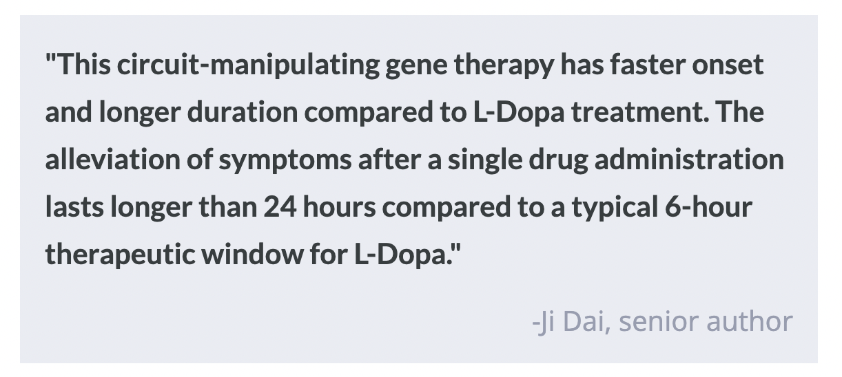
In a mouse model of PD, using their viral strategy to activate D1 striatal neurons resulted in a partial rescue of Parkinson-like motor deficits. The same strategy used in macaques with a model of PD resulted in a reduction or total reversal of all PD symptoms including tremor, bradykinesia, and rigidity, without affecting motor behaviors in naïve monkeys. This relief of PD symptoms lasted for eight months with consistent use of the same dose of designer drug required to activate the chemogenetic receptor. When compared to ongoing treatment with Levodopa, a current treatment for human PD, the authors’ designer viral strategy resulted in a longer window of symptom relief after drug administration and did not result in dyskinesia, a common negative side effect of long-term Levodopa treatment. This demonstrates the long-term safety and efficacy of using this strategy to treat the core symptoms of PD in primates. 

What's the impact?

This study found that chemogenetic manipulation of striatal dopaminergic neurons with designer drugs can enhance their activity and alleviate PD symptoms, and has improved long-term efficacy over current PD treatments. This means there may be safe, effective ways to increase the activity of the neurons that are compromised in PD, resulting in long-term improvements to symptoms like tremor and slow movement. This is promising for the future of gene therapy treatments in humans that target specific brain circuitry affected in PD and other neurodegenerative diseases and may lead to a treatment alternative for those in later stages of PD.