Gray Matter Loss in Psychosis is Present in Brain Regions Connected by White Matter

Post by Lani Cupo

The takeaway

Grey matter changes associated with the psychosis spectrum occur in networks connected by white matter, with the hippocampus as a central hub connecting regions of gray matter loss. 

What's the science?

The psychosis spectrum, from early first episodes to chronic psychotic disorders like schizophrenia, is associated with gray matter abnormalities, especially atrophy, across the brain. It is yet unknown, however, what mechanism underlies these changes. This week in JAMA Psychiatry Chopra and colleagues demonstrate that gray matter changes are constrained to networks connected by white matter (axonal connections), identifying the hippocampus as a potential source spreading volume loss to different regions.

How did they do it?

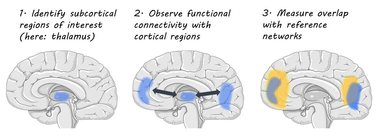
The authors acquired magnetic resonance imaging (MRI) data from 534 people in several groups: patients who had a first episode of psychosis (FEP), but no exposure to antipsychotics, patients who had been exposed to antipsychotics for less than three years, patients with established schizophrenia, and age-matched control groups for each patient group. Comparisons between each patient group and the corresponding control group were made to identify statistical differences in gray matter volume associated with psychotic disorders. In the FEP group, however, additional longitudinal analyses were conducted to isolate the effects of the disorder from those associated with antipsychotics. The authors examined change over time in a control group, patients receiving antipsychotics, and patients receiving a placebo. They could compare changes between the placebo group and control group to identify disorder-related changes and compare changes in the group that received antipsychotics to the placebo group to isolate antipsychotic-related changes. Using images that reflect which brain regions are connected (diffusion weighted imaging and functional MRI), the authors constructed a model of brain networks in a separate healthy dataset. They could then assess whether regions with volume change in the psychosis groups were part of the same brain networks using a coordinated deformation model. Next, the authors used a network diffusion model to assess whether changes in gray matter volume spread evenly throughout the network, or whether certain brain regions served as a hub, or epicenter for volume change.

What did they find?

First, the authors demonstrate that changes in gray matter across stages of illness are constrained by networks connected by white matter. Brain regions that were more strongly connected were more likely to show similar levels of gray matter loss. This suggests changes in various brain regions are not wholly independent, but rather connected in networks. In order to provide evidence that gray matter changes actually spread through axonal connections, however, the authors found that patterns of change associated with both illness and antipsychotic exposure were constrained by brain networks, implying that illness and medication-related changes over time are also related to the connectome. Finally, the authors identified the hippocampus as an epicentre for volume loss, as it was significantly different between patients and controls across all datasets.

What's the impact?

The results of this study suggest gray matter changes associated with psychosis may spread through axonal connections between regions. While there is little evidence for a protein-related spreading of psychosis-related pathology, the cellular profiles of connected regions may share alterations that underlie brain volume changes. Understanding the network-related changes may help future researchers identify the mechanism by which psychopathology impacts the brain to provide better treatment and prevention.

Neural Activity in Subcortical Regions is Highly Correlated with Resting State Cortical Network Dynamics

Post by Meredith McCarty

The takeaway

Despite the high interconnectedness of cortical and subcortical regions, the subcortex remains an understudied area of the human brain. High-resolution fMRI reveals that the subcortex is highly functionally connected (i.e. a high degree of correlated brain activity) with distinct cortical regions, demonstrating a gradient of connectivity for both integrative and segregated patterns of information processing.

What's the science?

In daily life, humans integrate information constantly, a process thought to be orchestrated by a highly interconnected brain. The brain is composed of many cortical and subcortical structures (i.e. neural formations deep within the brain), and there are complex patterns of connectivity between these regions that enable rapid network dynamics to unfold. To study these dynamics in healthy subjects, researchers can utilize noninvasive imaging techniques, although these methods are often limited in resolution to recording from the cortex. The lack of access to subcortical structures occurs primarily due to tooling and analysis limitations, including low signal-to-noise ratios and the varying properties of brain tissue in deeper structures. Because of these limitations, it is unknown how subcortical regions participate in neural information processing at a network level. This week in The Journal of Neuroscience, Groot and colleagues utilize high-resolution fMRI imaging techniques to measure the level of correlated activity between cortical and subcortical structures in humans.

How did they do it?

The authors utilized functional magnetic resonance imaging (fMRI) at a high-field resolution in order to record changes in BOLD signal across cortical and subcortical regions. They recruited 40 adults (21f) to participate in wakeful rest fMRI data collection, which consisted of two 15-minute sessions during which participants fixed their gaze on a central fixation point. They next determined cortical and subcortical regions of interest through use of automated parcellation algorithms. Their method of fMRI data analysis enabled the identification of patterns of intrinsic functional connectivity (FC) in the brain at a resolution that revealed more subtle functional organization. They compared subcortical FC patterns data with ‘reference networks’; common patterns of resting-state fMRI activation in the cortex known to be highly correlated with various behavioral and cognitive activities. These include the salience, visual, and default mode networks. By comparing the overlap between areas with FC to subcortical regions and the reference networks, the authors were able to measure the degree of network correlation between cortical and subcortical regions.

What did they find?

First, the authors found 7 subcortical regions that had high spatial correlation between brain regions they were functionally connected to and reference networks. These regions were the Thalamus, Striatum, Claustrum, Globus pallidus external, Hippocampus, Ventral tegmental area, and Substantia nigra. The authors dub the patterns of network correlation discovered to be “echoes” of intrinsic connectivity networks within cortical regions. Upon closer examination, they found a heterogeneous organization of echoes within subcortical subregions, with some regions exhibiting high correlation with many reference networks, while other regions exhibited very little to no significant connectivity. This suggests that there is a gradient within subcortical regions of distinct and integrated network dynamics.

What's the impact?

This study found activity in distinct subcortical regions to be highly correlated with distinct resting state cortical networks. These results suggest that the subcortex is involved in integrated multi-network neural activity with many cortical regions, an area of research previously restricted by methodological constraints. Altered subcortical dynamics are linked to many brain disorders, therefore a greater understanding of the role of subcortical regions in resting state healthy brain dynamics is pivotal.

Access the original scientific publication here

Learning Fear Extinction is Driven by Dopamine Neurons in the Midbrain

Post by Rebecca Hill

The takeaway

Dopamine neurons help signal fear extinction - the ability to stop fear responses to stimuli that are no longer dangerous. Neuronal pathways in the midbrain are required for dopamine signaling in fear extinction learning.

What's the science?

In order to adapt to signals that are no longer dangerous, animals must learn how to stop fear responses through fear extinction learning. Individuals with post-traumatic stress disorder (PTSD) also require this ability to inhibit fear responses. While previous research has shown the involvement of dopamine neurons in learning fear extinction, it is still unclear which specific neuronal pathways are involved. This week in Neuron, Salinas-Hernandez and colleagues identified these dopamine pathways in the midbrain by measuring dopamine neuron activity.

How did they do it?

In order to learn to associate signals with new and different outcomes (dangerous or not dangerous), animals must use prediction errors: where a prediction is met with a different result, an error. To capture brain activity associated with these prediction errors, the authors used fiber photometry to measure activity-dependent calcium signals from dopamine neurons while mice underwent fear extinction paradigms. This involved first playing mice a tone to habituate them to the signal. Next, the mice are conditioned to fear the tone, by pairing it with a foot shock. Last, they played the tone by itself for two days to create the extinction of the fear response. The authors measured dopamine neuron activity in the midbrain of mice both before the fear conditioning and after. They also measured the response to positive signals by rewarding mice with sugar water for nose-poking a target area. To observe whether the fear extinction response was dopamine neuron dependent, the authors optogenetically inhibited dopamine neurons in NAc. Finally, the authors used chemogenetic inhibition to test whether neurons in specific areas of the midbrain were used to signal for prediction error signals.

What did they find?

The authors found that dopamine neurons showed more activity in certain areas of the midbrain — the ventral tegmental area (VTA) and the nucleus accumbens (NAc) — during extinction than during habituation. This suggests these pathways of dopamine learning are used in fear extinction learning. Specifically, neurons in the anteromedial NAc responded with more activity than in other areas. This suggests the prediction error signal is carried to this specific area of the NAc. The authors also saw more calcium signaling, and higher activity, when mice were rewarded by sugar water, which suggests these areas also are activated by rewards. Finally, when dopamine neurons were inhibited in NAc, mice were less able to exhibit extinction learning and continued to display a fear response to just the tone. This provides further evidence that dopaminergic neurons projecting to NAc control fear extinction. Finally, when the authors inhibited dopamine neurons that projected to the VTA (a region with neurons that project to the NAc), mice could not learn fear extinction, suggesting that VTA-projecting dopamine neurons are required for prediction error generation.

What's the impact?

This study is the first to show that midbrain regions control the fear extinction learning for dopamine neurons. This work furthers our understanding of brain regions involved in fear extinction learning and could have future implications in humans. More broadly, it could point to possible treatments for disorders such as PTSD.

Access the original scientific publication here.