The Cytokine IL-12 Protects Against Neuroinflammation by Signaling to Neurons

Post by Trisha Vaidyanathan

The takeaway

The cytokine interleukin-12 (IL-12) has a protective effect in neuroinflammatory diseases. The neuroprotective effect of IL-12 is mediated by neuronal sensing of IL-12 via the IL-12 receptor, resulting in transcriptional changes that increase survival, proliferation, and protection.

What's the science?

Central nervous system autoimmune disorders like multiple sclerosis (MS) are characterized by cytokine dysregulation, in which the body’s own immune system attacks tissue, causing inflammation that ultimately damages the brain. The cytokine IL-12 is a well-established driver of this type of inflammation. Paradoxically, mice lacking IL-12 develop worse neuroinflammation and clinical outcomes in several disease models, suggesting IL-12 also has a protective role. This week in Nature Neuroscience, Andreadou, Ingelfinger, and colleagues address this paradox by investigating the hypothesis that IL-12 elicits different effects depending on the cell type receiving the signal.

How did they do it?

First, the authors asked which cells are necessary to mediate the neuroprotective effect of IL-12. Using the cre-lox system, the authors were able to delete the IL-12 receptor from specific cell populations in a mouse model of MS (the EAE model) and investigate which cell-type specific deletion altered clinical outcomes.

Second, the authors used immunohistochemistry and RNA-fluorescence in situ hybridization to visualize IL-12 receptors and IL-12 receptor mRNA transcripts, which allowed them to identify which cell types in the brain are capable of sensing IL-12. Additionally, the authors confirmed their findings from the mouse in humans by analyzing human RNA-sequencing datasets and performing immunohistochemistry in MS patient tissue.

Third, the authors performed single nuclei RNA sequencing on mice where the IL-12 receptor was deleted in neurons and oligodendrocytes in order to reveal several cell-type specific transcriptional changes induced by the absence of the IL-12 receptor. Lastly, the authors used a neuronal cell culture system to test the effect of IL-12 on isolated mouse neurons and confirm their findings on the role of neuronal IL-12 signaling in neuroprotection.

What did they find?

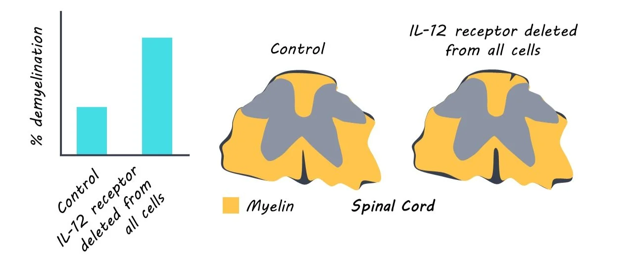
First, the authors deleted the IL-12 receptor from all cells and found the mice exhibited worse MS clinical symptoms, confirming previous findings that IL-12 can be protective. Next, the authors demonstrated that the protective effect was not driven by the sensing of IL-12 in immune cells because deleting the IL-12 receptor in all bone-marrow-derived immune cells, just T-cells, or just natural killer (NK) cells, did not change clinical disease symptoms in the mice.

Second, the authors demonstrated that both neurons and oligodendrocytes in the mouse brain express the IL-12 receptor. These findings were also confirmed in human MS patient tissue and sequencing datasets. Deleting the IL-12 receptor in neurons and oligodendrocytes led to worse disease symptoms and neuroinflammation, revealing that the neuroprotective effect was dependent on these cells.

Third, the authors answered the question of what IL-12 signaling in the brain does. The deletion of the IL-12 receptor in neurons and oligodendrocytes led to several transcriptional changes across many cell types. Overall, they found a reduction in gene expression for neuroprotection and survival, as well as a broad class of proteins called trophic factors released by cells to protect and support other cells.

Lastly, the authors narrowed in to identify that neuroprotection is mediated by neurons, not oligodendrocytes: When the authors deleted the IL-12 receptor only in oligodendrocytes there was no change in the MS clinical symptoms. To confirm this, the authors administered IL-12 to a cell culture system where only mouse neurons were present and found IL-12 increased the expression of genes that promote neuroprotection and remyelination as well as induced the release of trophic factors that are important for oligodendrocyte survival and function.

What's the impact?

This study is the first to settle the paradox of IL-12 by demonstrating that IL-12, a well-established driver of inflammation, can protect against inflammation by signaling to neurons. Understanding the protective effect of IL-12 can provide critical insight for clinical interventions in autoimmune disorders or neurodegenerative diseases that cause neuroinflammation. 

Modelling Human Psychological Responses to Robots: The Positive, the Negative, and the Competent

Post by Rachel Sharp

The takeaway

As robots become increasingly present in our everyday lives, human reactions to them become more complex. Human responses to robots can be categorized into three dimensions – positive, negative, and competence-related, and predictors can be identified for each response category, thereby establishing the positive-negative-competence (PNC) model.

What's the science?

Robots are becoming increasingly more integrated with human society, resulting in more common and diverse human-robot interactions. Understanding how we think, feel, and react to robots in different spaces informs how we incorporate robots into pre-existing social structures. Previous studies on this topic have not included a comprehensive framework for the range of psychological responses we have, nor the diverse types of robots that humans interact with today. This week in Nature Human Behavior, Krpan, Booth and Damien sought to develop a model that could classify the broad spectrum of psychological responses to robots, organize these responses into grouped patterns (dimensions), and identify thought patterns that most strongly predicted these dimensions.

How did they do it?

The authors conducted 7 studies over 3 phases to develop a framework of robots (Phase 1; studies 1 & 2), develop a categorization for how we respond to robots (Phase 2, studies 3-5), and determine what best predicts these responses and why (Phase 3, studies 6 & 7). Across studies, participants responded to stimuli of robots as images and descriptions across 28 areas of human activity where robots are present. First, a group of participants were asked to generate characteristics they associated with robots, resulting in 277 unique characteristics. Then, a new set of participants grouped these characteristics into categories, from which 5 clusters of robot characteristics were found using hierarchical clustering. The authors then linked the themes of these clusters, forming a general definition of robots. Participants were then given this definition and asked to list all human domains they could think of that robots operate within. A new set of participants were given this comprehensive list of domains and asked to list all thoughts, feelings, and behaviors they could think of regarding robots operating in these domains. Based on their responses, the authors generated prompts to probe the reported thoughts and feelings, and asked new participants to answer them in response to an example of a robot from one of the original 28 domains (i.e. “This robot is like a human”). Responses to these prompts were used to generate statistical models to measure underlying factors representing the diversity of responses.

They found that responses could be most appropriately represented by 3 categories: positive, negative, and competence-related. They then confirmed that the model remains accurate across changes in participant demographic information or robot type. Lastly, the authors trained machine learning models for the positive, negative, and competence dimensions separately and identified the key predictors for each response type. They validated the identified predictors and used parallel mediation analysis to analyze potential mediators of the relationship between a predictor and the associated PNC dimension. For example, general risk propensity was a predictor for the positive dimension because people who scored highly for this trait valued the risks associated with using robots in society and were curious about how they would influence the world.  

What did they find?

The authors established the positive-negative-competence (PNC) model to represent the spread of human psychological responses to robot representation. They also identified unique predictors for each of these dimensions and were able to identify mediators for 3/3 of the positive dimension predictors, 2/4 of the negative dimension predictors, and 2/2 of the competence dimension predictors. The key positive predictors of each dimension with an example of an identified mediator of the predictor in parenthesis, where applicable, were: general risk propensity (valued risks of robot use), anthropomorphism (felt positively towards inanimate objects with human features), and parental expectations (felt robots could help humans fulfill their high expectations). The key predictors for the negative dimension were trait negative affect (were more likely to be in a state of displeasure), psychopathy (felt inferior towards technologies they were not proficient in), anthropomorphism (no mediator found), and expressive suppression (no mediator found). For the competence dimension, key predictors were approach temperament (valued exceptional skills and competencies) and security-societal (linked advanced technologies with the degree of societal advancement). 

What's the impact?

While previous research on human-robot relations has examined psychological responses to robots, this is the first study to investigate the spectrum of human psychological reactions under a comprehensive construct. Importantly, the authors illustrated that while the spectrum of human responses to robots is diverse, they can be explained by three dimensions of psychological processing: positive, negative, and competence-related. The proposed model allows future research to measure responses to robots more easily and accurately, helping us to construct a framework to understand how humans think, feel, and react to robots encountered in society.

The Role of Extracellular Potassium in Regulating Wakefulness

Post by Christopher Chen

The takeaway

Extracellular potassium ([K+]e) in the brain has long been linked to arousal states. Researchers demonstrate that potassium dynamics affect neuromodulator release and impact cortical activation, suggesting that potassium may influence phenomena like local sleep during wakefulness and abnormal sleep/wake cycles following brain injuries.

What's the science?

Our brains cycle through various states of alertness over the course of the day. For example, when we sleep our brains experience high-amplitude slow-wave activity, representing the synchronized firing and inactivity of large neuron populations. Conversely, during wakefulness, our brains experience low-amplitude high-wave activity characterized by desynchronized firing and elevated neuronal activity. Monoamine neurotransmitters, or neuromodulators, are closely tied to changes in brain activity patterns and arousal levels. 

In a region of the brain called the locus coeruleus (LC), a subset of monoaminergic neurons that release neuromodulators like norepinephrine (NE) and dopamine (DA) project to the cortex and show different discharge rates during wakefulness, nonrapid eye movement (NREM) sleep, and rapid eye movement (REM) sleep. Changes in cortical potassium levels have also been linked to arousal, particularly the sleep-wake cycle. When we sleep, potassium levels are low ~(3 mM) but gradually increase to ~4 mM as we enter a wakeful state. Despite the role of potassium in arousal, researchers have not extensively studied how directly manipulating potassium alters NE levels and behavioral states. 

Recently in PNAS, Dietz et al. manipulated potassium levels in mice to measure its effects on LC activity and NE levels as well as its overall impact on the brain. In doing so, they provide a new perspective on how our brains undergo behavioral state changes and suggest that potassium may have an underappreciated role in cortical function. 

How did they do it?

In order to manipulate potassium levels as well as measure NE levels and the levels of other well-known neuromodulators like DA and serotonin (5-HT), the researchers used a process called in vivo microdialysis that allowed them to inject various concentrations of potassium into the brains of mice, and a process called high-performance liquid chromatography (HPLC) to measure the levels of potassium and neuromodulators. 

To test how potassium changes affected arousal they utilized a tail shock to induce arousal in the mice and measured how this arousal response differed in conditions with various potassium levels. They also measured how changes in potassium levels affected sleep/wake cycles by comparing brain activity in mice implanted with EEG electrodes. Finally, as disturbances in the sleep/wake cycle have been shown to affect physical activity and performance on motor tasks, researchers measured how changes in potassium levels influenced wheel running and motor performance.    

What did they find?

To test potassium-dependent effects on neuromodulator levels, researchers injected various concentrations (2.5, 3.5, and 5 mM) of potassium into the brain during the light phase and dark phase of the day. They discovered that in both phases, increasing potassium levels induced parallel increases in NE, DA, and 5-HT and decreasing potassium levels resulted in parallel decreases in NE, DA, and 5-HT.

Having established the direct effects of changes in potassium levels on neuromodulators, researchers tested whether these potassium changes influenced arousal. Following the injection of various potassium levels, they discovered that injecting low levels of potassium prior to a tail shock limited the mouse’s response as measured by NE levels. In fact, these mice expressed almost no tail shock-induced increase in NE. Conversely, NE levels in mice injected with high (5 mM) potassium were nearly 2x as high as the NE levels induced by a baseline startle response, highlighting the influence of potassium on arousal state.

In terms of the sleep/wake cycle, mice injected with 2.5 mM spent significantly more time in NREM and REM sleep and significantly less time in the wake state as measured by EEG activity. These mice were also less active than the control group, expressing less overall spontaneous movement.

Lastly, the researchers sought to measure the effects of potassium changes on wheel running and motor performance. Interestingly, increasing potassium levels correlated with further distance traveled on a wheel but did not significantly affect the number of times the mice used the wheel, suggesting that potassium levels influence the sustainment of running. As for motor performance, increases in potassium levels directly correlated with decreases in the number of falls from a rotarod, suggesting high levels of potassium contributed to heightened motor performance.  

What's the impact?

While changes in potassium levels have been linked to changes in brain function, this study provides a detailed explanation of how direct manipulations of in vivo potassium levels in mice alter the brain as well as discrete behaviors. Clinically, researchers may rely on these findings to inform therapeutic approaches for aberrant sleep/wake cycles caused by brain injury. Finally, it provides evidence that a local (cortical) source in the form of potassium may be influencing local NE release, a novel finding that may potentially reconfigure our understanding of how arousal is processed in the brain.