A “Pro-Inflammatory” Diet is Associated with Increased Inflammation and Cognitive Decline

Post by Lani Cupo

The takeaway

A diet high in pro-inflammatory foods is associated with proteins related to inflammation in the blood and later-life cognitive impairment (associated with Alzheimer’s Disease [AD]) in a large cohort of caucasian women.

What's the science?

Previous studies have linked certain dietary patterns with elevated markers of inflammation. Additionally, increased inflammation has been associated with cognitive decline in aging. It is unknown, however, how diet relates to a wide array of inflammatory markers, and which of these play a role in cognitive decline in aging. This week in Molecular Psychiatry, Duggan and colleagues explore the relationship between dietary patterns, markers of inflammation, and cognitive decline in a large group of women over time.

How did they do it?

The authors used a subset of data from the Women’s Health Initiative Memory Study which includes samples from 1528 women, on average aged 71 at the first timepoint. At the first timepoint, researchers surveyed participants about their diet, collected blood samples to assess markers of inflammation, and performed assessments of cognition. Each annual follow-up included cognitive assessments as well. Using a pre-defined tool known as the Dietary Inflammatory Index, the authors placed each diet on a continuum from “anti-inflammatory” (e.g. tomatoes, fruits, nuts) to “pro-inflammatory” (refined carbohydrates, fried foods). 151 inflammatory and immune proteins were assessed in blood samples and included for analysis. Cognition was assessed by clinicians, who categorized participants as having “no impairment”, “mild cognitive impairment”, or a “probable dementia.” Importantly, the authors controlled for covariates that could confound the relationship between an inflammatory diet and cognitive decline, such as education. Finally, in separate datasets, the authors validated the association of proteins identified in the first study with a) age to onset of dementia in both sexes and b) brain atrophy measured with magnetic resonance imaging in both sexes in areas related to AD.

What did they find?

First, the authors found in the main sample that a more inflammatory diet at baseline was associated with 55 of the 151 inflammatory proteins in blood samples, including, for example, proteins involved in pro-inflammatory signaling (interleukin-6), and regulation of phagocytosis. They also found associations between an inflammatory diet and genes regulating inflammatory responses and responses to pro-inflammatory signals. Of the proteins associated with an inflammatory diet, the authors found 6 proteins were weakly associated with an increased likelihood of future cognitive impairment, with these proteins involved in processes such as gene transcription and immune signaling. In the first validation study, 5 of the 6 proteins had been measured, and all 5 of them were associated with increased risk of dementia and 3 of them were associated with earlier onset of dementia. From the second validation study, the authors found 3 of the 6 proteins were associated with greater brain atrophy in regions associated with AD-related dementia, and all 3 were also associated with increased rates of dementia in the first validation study.

What's the impact?

This study found a set of proteins that were associated with an inflammatory diet, a subset of which were also associated with cognitive impairment, the risk for AD, and AD-related brain atrophy. The genes that regulate these proteins could become possible therapeutic targets for AD treatments. An anti-inflammatory diet could also provide a protective effect for high-risk individuals, although further research is needed to confirm this.

Access the original scientific publication here.

Human Brain Organoids Can Integrate into the Adult Rat Visual System

Post by Megan McCullough

The takeaway

Human brain organoids, grown from human stem cells, can integrate both structurally and functionally into adult rat brains.

What's the science?

One promising treatment for restoring brain function after an injury is cell transplantation. Human brain organoids, created from pluripotent stem cells, are an avenue of research currently being studied for therapeutic potential. Previous studies have shown that it is feasible for human brain organoids to integrate into rat brain systems, but there has been limited research into the functional integration of these organoids into the networks of injured mammalian brains. This week in Cell Stem Cell, Jgamadze and colleagues transplanted human brain organoids into damaged cavities in the visual cortex of adult rats to examine potential integration. 

How did they do it?

The authors transplanted brain organoids into the visual cortexes of rats. These tissues were generated from human stem cells and expressed the fluorescent marker GFP - a protein that lights up once exposed to ultraviolet light, allowing the authors to identify the organoids once grafted into the rat brains. The organoids had been growing for around 80 days, longer than in previous studies, to allow for maturation and cell differentiation. The authors then conducted a histological (tissue) analysis on the human grafts to confirm the human origin of the cell tissues, examine potential cell maturation, and observe any immune response from the host tissue. This was done to examine how the cell grafts evolved over time. Viral tracers were also injected into the eyes of the rats which allowed the authors to observe the connections between the human cells and rat cells. Next, the authors looked for functional integration of the transplanted organoids. Extracellular recordings were conducted to test for neural activity in the grafted tissue in response to presenting the rats with visual stimuli.

What did they find?

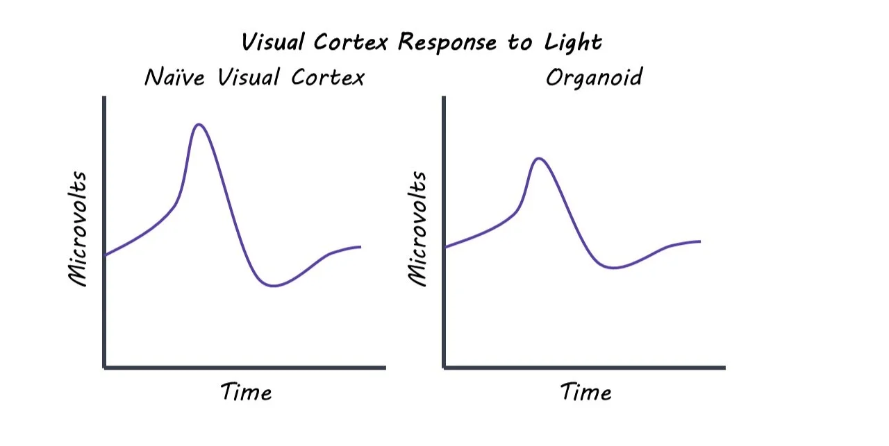
The authors found that in a three-month time period, human brain organoids integrated into the visual system of rat brains. Histological analysis showed that although there was inflammation at the graft site, there was only a mild immune response from the host tissue. This suggests that cell transplant can be a viable option into mammalian brains. This analysis also showed that the human cell tissue continued to differentiate and mature once transplanted. In addition to structural integration, the transplanted organoids also showed functional integration. The grafted human neurons received inputs from the visual system, forming connections via synapses with the host neurons. The brain organoids demonstrated both spontaneous and evoked neural activity, providing further evidence of functional integration. When the rats were exposed to flashing lights, the organoid neurons responded to the stimulation in similar way that the host cells responded.  

What's the impact?

This study found that human brain organoids can both structurally and functionally integrate with the visual system of rats. The human neurons formed connections with the rat neurons and displayed electrical activity in response to visual stimulation. This research shows the possibility of using lab-grown neural tissue to reconstruct brain circuitry after brain injury or stroke. 

CBD’s Anti-Seizure Mechanism of Action

Post by Baldomero B. Ramirez Cantu

The takeaway

Hippocampal hyperactivity is a hallmark of a variety of neurological diseases. Cannabidiol (CBD) is capable of modulating the excitatory-inhibitory balance of hippocampal neurons and its effects are mediated by interactions with the GPR55 receptor.

What's the science?

CBD is known to reduce seizure activity in animal models and patients with treatment-resistant epilepsies. However, the underlying mechanisms that enable CBD to reduce seizure activity and severity remain unknown. This week in Neuron, Rosenberg and colleagues investigate the role of the GPR55 receptor and its endogenous ligand LPI in CBD’s anti-seizure action.

How did they do it?

The authors tested if the GPR55 receptor is the site of action for CBD's anti-seizure effects by using mice with functional or genetically-removed GPR55 receptors. They conducted experiments on hippocampal neurons in-vitro to study how GPR55’s natural ligand, LPI, affects neuronal excitability and how CBD interacts with LPI’s effects. Finally, to investigate changes in GPR55 receptor expression after seizures in control or CBD pre-treatment conditions, the authors used qPCR and immunohistochemistry. Seizures were induced in the mice using the drug pentylenetetrazole or lithium-pilocarpine.

What did they find?

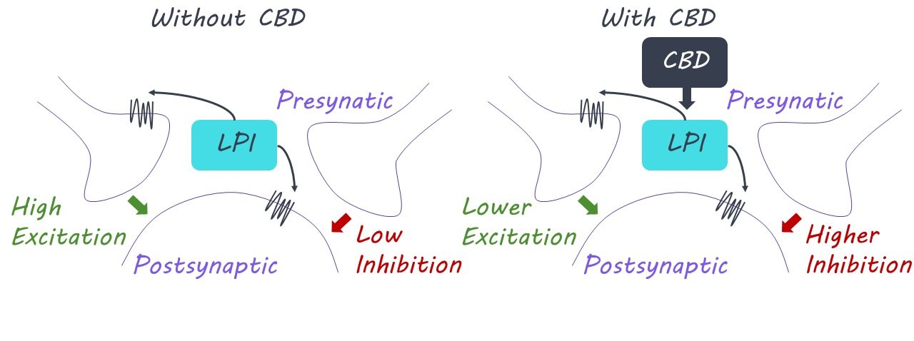
The authors found that seizures were only reduced by CBD if mice expressed GPR55 receptors - there was no improvement if these receptors were removed. This finding suggested a critical role of the GPR55 receptor in enabling CBD to attenuate seizures. They conducted neural recordings from the hippocampus and found that the application of LPI increased neuronal excitability in neurons from normal mice, but not in those from genetically modified mice lacking GPR55 receptors, indicating that LPI drives GPR55-dependent increases in neuronal excitation. LPI was found to disrupt neuronal activity in several ways, such as interfering with the impact of crucial inhibitory interneurons that are essential for maintaining an appropriate excitatory-inhibitory balance in the hippocampus. Importantly, hippocampal neural recordings showed that the presence of CBD counteracts the increased excitability produced by LPI. This provides evidence for the GPR55 receptor as a functional target of CBD’s anti-seizure action. Mice that received CBD pre-treatment showed significantly lower increases in GPR55 immunoreactivity and receptor mRNA expression after induced seizures compared to the control group.

What's the impact?

This is the first study to identify the two-pronged molecular signaling, affecting both excitation and inhibition, underlying CBD’s anti-seizure action. These findings could serve as the foundation for the development of new, targeted anti-epileptic therapies