The Effect of Bilingualism on Subcortical Brain Structures

Post by Megan McCullough

The takeaway

Speaking two languages is a cognitively demanding task that has a measurable impact on the volumes of subcortical brain structures. However, this relationship can be non-linear and reaches a plateau after a certain level of bilingual experience has been reached.

What's the science?

Bilingualism, the ability to fluently speak two languages, is a complex cognitive skill. Previous research has demonstrated that engaging in cognitively demanding tasks can increase grey matter volumes in the brain, but this increase is nonlinear, plateauing or even decreasing after a certain point. The expansion-renormalization model of experience-dependent neuroplasticity explains these findings. However, so far it is unclear whether these nonlinear changes apply also to bilingualism. In a recent study published in Scientific Reports, Korenar, and colleagues investigated the impact of bilingual experiences on subcortical brain regions involved in cognitive control and language selection, specifically the basal ganglia and thalamus.

How did they do it?

Participants in this study were native Slavic language speakers who had a good command of English. Bilingualism is not a one-size-fits-all phenomenon, so the study did not consider participants as bilingual just because they spoke two languages. Instead, each participant was given a composite score to reflect the richness of their individual bilingual experience. The authors effectively treated bilingualism as a continuum of experiences, which allowed them to study the relationship between bilingual experience and changes in subcortical structure volume. Magnetic resonance imaging was used to measure the volume of the caudate nucleus, globus pallidus, putamen, nucleus accumbens, and thalamus. These structures are important for controlling behavior and managing two languages in the brain. The authors then used non-linear statistical modeling, specifically generalized additive mixed models (GAMMs), to investigate whether bilingual experiences had an impact on subcortical volumes that were consistent with the model of experience-dependent neuroplasticity. By using GAMMs, the authors were able to model the possible non-linear effects of bilingual experience on subcortical volumes.

What did they find?

The authors of the study have discovered that bilingualism has a measurable impact on the volume of subcortical structures such as the basal ganglia and thalamus. Interestingly, this relationship is non-linear and dependent on the bilingual composite score, which measures the level of bilingual experience. Specifically, there is a positive association between the bilingual composite score and the volumes of the caudate nucleus and the nucleus accumbens. However, this relationship reaches a plateau in individuals with extensive bilingual experience, indicating a renormalization process. The Dynamic Restructuring Model provides a possible explanation for these findings. According to this model, acquiring and using an additional language leads to an expansion of brain structures as new neural pathways are formed. However, this expansion is followed by a contraction as less efficient pathways are pruned, leaving only the most efficient connections. This suggests that becoming more proficient in handling two languages allows the brain to selectively keep only the most efficient neural pathways for this task. The study's data suggest that the bilingual composite score can be used as a predictor of subcortical structure volumes important for using two languages. Moreover, the study shows that bilingualism impacts subcortical structure volumes in a manner similar to other cognitively demanding tasks previously studied, like playing a musical instrument.

What's the impact?

Overall, this study is the first to investigate the non-linear relationship between bilingual experiences, volumes of subcortical brain regions, and a continuous measure of bilingualism. The findings suggest that acquiring and speaking a new language can prompt the brain to grow and shrink, as a function of efficiency, similar to other cognitively demanding tasks.

Brain Recording and Stimulation— On the Go!

Post by Anastasia Sares

The takeaway

Researchers have designed a compact, portable device that can record brain activity from patients who already have electrodes implanted in their brain for medical reasons. The expanded capabilities of this device will allow us to test more complex forms of clinical neuromodulation, as well as provide a view into the neuron-level functioning of the human brain in life-like situations.

What's the science?

The number of people with therapeutic electrodes implanted in their brains, though small, is rising. The classic use of this technology is with epilepsy patients, to monitor a region suspected of generating seizures before operating. Nowadays, more permanent devices are being tested which could detect the beginnings of a seizure and deliver a pulse of electricity to stop it in its tracks. Another reason someone might have electrodes implanted is for deep brain stimulation (DBS), which can be used as a treatment for Parkinson’s disease and depression, among other disorders. In addition to their clinical applications, these electrodes are a unique opportunity for researchers to measure human brain activity at the single neuron level.

This week in the journal Nature Neuroscience, Topalovic and colleagues present their new system, Neuro-stack, a wearable device that can connect to a patient’s already-implanted brain electrodes, recording activity and also stimulating particular brain regions.

How did they do it?

The Neuro-stack combines many existing technological elements important to both research and clinical applications, while also increasing their resolution and power. Some features include: 

  • Portability: people can wear the device during an experiment and not be hampered by wires tethering them to computers or other devices.

  • Simultaneous recording of single-neuron activity and fluctuations of entire neuron populations (known as local field potentials or “LFPs”)

  • Precise electrical stimulation (sending a pulse of electricity into the brain at a targeted location)

  • Correction: Statistical techniques to account for electrical stimulations so they don’t interfere with recordings.

  • Neural networks that can learn what brain activity is relevant for a given task 

  • Eye tracking and a world-view camera, so information from a person’s environment can be synchronized with their brain activity.

What did they find?

The team demonstrated the capabilities of the Neuro-stack in a few small experiments. In one, participants walked around the room while the device recorded activity in the neurons of a region called the hippocampus. Just like in previous experiments with rodents, the hippocampus responded to the person’s location in the room, firing more often when they were close to the walls. This confirms that humans have some of the same navigation mechanisms as rodents when it comes to spatial boundaries.

Another validation experiment was a memory test. In each block, a participant was given a list of ten words to memorize. They then completed a math test as a distracter and finally were asked to recall the words at the end of the block. The authors used a machine learning algorithm (convolutional and recurrent neural networks) to predict which word would be remembered and which would be forgotten based on the activity in the brain at the time of hearing the word.

These experiments were small, proof-of-concept studies and studies with more people would be needed to fully flesh out the findings. However, these small demonstrations show that the Neuro-stack is a useful tool and works as intended.

What's the impact?

Portable implanted electrode systems are often used in animal research and have led to many discoveries about how the brain works. However, a rodent and a human still have many differences, and a rodent cannot converse with a researcher or give feedback about their experiences using language. In addition, certain psychological conditions are difficult to replicate or detect in rodents, limiting clinical research. The Neuro-stack could help to bridge the gap between animal and human research, increasing our understanding of the human brain. Of course, we will have to continue to be vigilant of the ethical implications as our digital and biological worlds collide.

Aggression-Specific Mirror Neurons Exist in the Hypothalamus

Post by Lincoln Tracy

The takeaway

A subset of neurons essential for aggression in the ventromedial hypothalamus of the mouse is activated when observing aggression in other animals, providing an example of mirror neurons for this specific social behavior. 

What's the science?

Mirror neurons are a subtype of neurons activated both during seeing and undertaking a behavior. Consequently, they have been suggested to be an important component of the cognitive substrates underlying successful social interactions. Activating neurons in the ventrolateral section of the ventromedial hypothalamus – the ‘attack’ center of the brain – elicits aggression in mice, but it’s unknown whether these neurons are also activated when observing aggression between other animals. This week in Cell, Yang and colleagues used calcium imaging and multiple types of genetically modified mice to determine whether neurons in the ventrolateral section of the ventromedial hypothalamus could perceive aggressive encounters between other mice.

How did they do it?

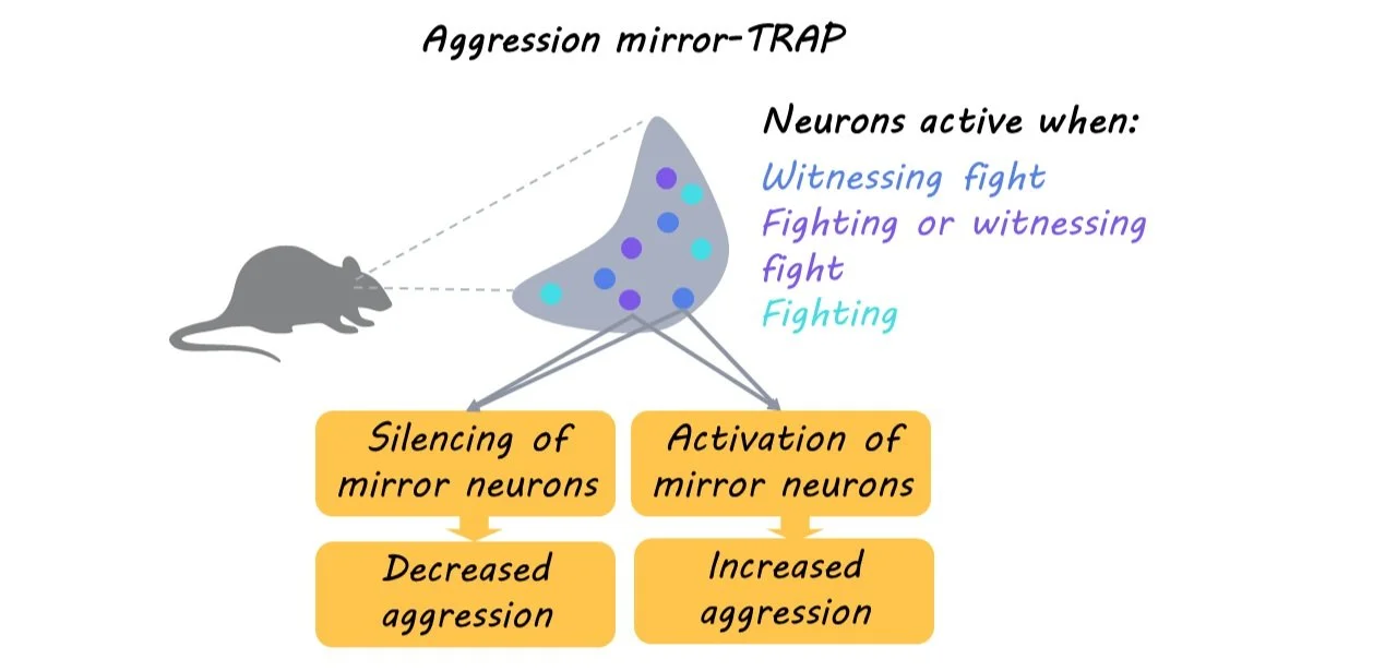
First, the authors used fiber photometry to determine whether aggressor neurons in the ventromedial hypothalamus of male mice were activated in response to social interactions with a male intruder in their cage. Second, they tested whether observing, but not participating in, aggression between two other mice activated the neurons in a similar fashion by separating the mice with clear, perforated plastic partition. Third, they tested which specific aspects of observing aggression were required for neuronal activation by altering the mice’s ability to smell and see the other animals. Fourth, they used socially naïve and experienced males to determine whether aggression mirroring requires the mouse to have previously participated in aggressive behaviors. Fifth, they used a TRAP2 mouse model to specifically test whether neurons activated in observers were also activated when the mouse participated in aggressive behaviors. Finally, they tested if the aggression-mirroring neurons were functionally relevant for fighting by using an aggression mirror-TRAP (aggression mirror neurons tagged with TRAP) to inhibit these neurons in a territorial aggression paradigm. 

What did they find?

First, the authors found aggressor neurons in the ventromedial hypothalamus of male mice were activated when a mouse interacted with and attacked a male intruder that had been placed in its cage. Second, just as when the mouse participated in aggressive behaviors, observing other mice be aggressive resulted in similar activation of ventromedial hypothalamic neurons. Third, they found that visual input is necessary for the activation of aggression-mirroring hypothalamic neurons, as repeating the experiment under infrared light did not activate aggressor neurons. Fourth, they found prior experience was not needed, as ventromedial hypothalamic activation was similar for both socially naïve and experienced mice when observing aggressive behaviors between other mice. Fifth, they found that there was an overlap of ventromedial hypothalamic neurons that were activated in observers and participants of aggression. Finally, they found the aggression mirroring neurons were essential for territorial fighting, as forced inhibition of these neurons reduced aggressive, but not non-aggressive social, behaviors. In additional studies, they also found that activating aggression mirroring neurons was sufficient to elicit aggression.

What's the impact?

These findings provide a genetic platform that can help us gain molecular and cellular insights into how individual neurons represent social behavior like aggression with respect to both mirroring and performing actions.