Finding Better Ways to Target Epilepsy Zones in the Brain

Post by Christopher Chen

The takeaway

Surgically removing the epileptogenic zone (EZ) – the brain region where epileptic seizures are generated – can allow patients with drug-resistant epilepsy (DRE) to live seizure-free, but it is often challenging to locate the EZ’s precise location. By mapping the propagation of interictal spikes - brief spontaneous neural discharge between seizures - researchers discovered key components of successful surgical resections that may help improve health outcomes in patients with DRE.

What's the science?

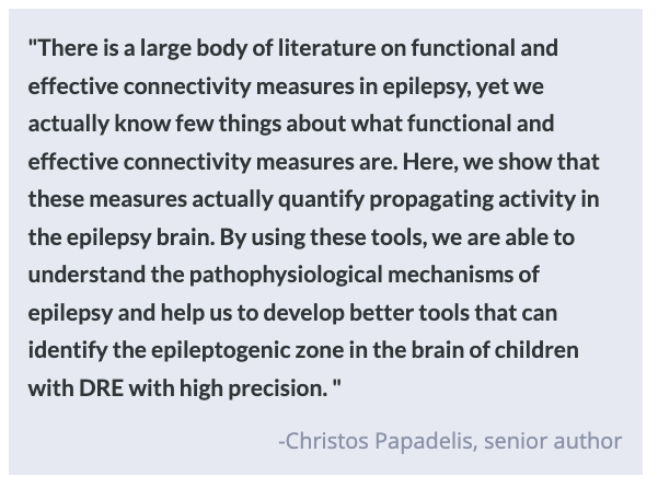
In patients with drug-resistant epilepsy (DRE), complete surgical removal of the epileptogenic zone (EZ) – the brain region where seizure activity is generated – can allow patients to live a life free from seizures. Currently, clinicians locate the EZ using a biomarker called the seizure onset zone (SOZ) which is identified by using a technique called intracranial electroencephalography (iEEG). However, iEEG has several technical limitations that may lead to defining a SOZ that does not encompass the entire EZ, thus leading to incomplete EZ resection and poor clinical outcomes.

To offset these limitations, clinical researchers have devised a new technique combining electric source imaging (ESI) with iEEG to provide more precise identification of the EZ. ESI allows for the mapping of propagating electric signals following a seizure (i.e., interictal spikes) and clearer delineation of brain function following a seizure. In a recent article in Brain, researchers used this strategy on patients with DRE to identify physical and network signatures that correlate with both good and poor surgical outcomes. 

How did they do it?

To characterize brain activity in patients with both good and poor surgical outcomes, researchers retroactively analyzed brain imaging and network activity data from roughly 40 children with DRE at a single hospital site in the United States over a six-year period. Working with a team of clinicians, researchers analyzed patient data to generate 3D images of each patient’s brain along with functional maps of brain activity.

Researchers then needed to characterize the physiological and network characteristics of patient brains. They used ESI to help reconstruct three key spatiotemporal zones of interictal spike propagation: spike onset, early spread, and late spread. In other words, they measured and identified the physical location of the spike signal in the brain over the first 10% of its spread (onset), the next 10% of its spread (early spread), and the final 80% of its spread (late spread). Researchers also measured the level of functional connectivity between these three zones as well as their degree of physical overlap with the surgically-resected region. These data points were then compiled and analyzed in order to compare profiles of patients who had good and poor outcomes from surgical resection.  

What did they find?

There were several notable findings pertaining to the patient profiles from good and poor surgical outcomes. In terms of the type of electric events, there were no major differences in the type of spike activity (isolated vs. propagating) and only slight differences in the descriptive characteristics of the spikes themselves (e.g. spike velocity) between patient groups. However, there was a key difference in the actual direction of the spike spread. Specifically, spike spread was more organized and hierarchical in patients with good outcomes (onset -> early spread -> late spread). Researchers also found that patients with good outcomes had surgical resections physically closer to the onset, early and late spread zones. 

Furthermore, when researchers delved deeper into the patient profiles with good outcomes, they found that their surgical resections were closest to one zone in particular, the spike onset zone and that surgical resection of the spike onset zone was an accurate predictor of surgical outcome. Interestingly, researchers also found that resection of the clinically-defined SOZ was not an effective predictor of good surgical outcomes. Overall, these findings point to the idea that patient profiles with good surgical outcomes were highlighted by more directionally-organized information flow and resection that was close to the spike onset zone.

What's the impact?

This research highlights network connectivity signatures and spike propagation analysis as critical strategies in identifying the EZ in patients with DRE. Researchers were able to link resection of the spike onset zone to good surgical outcomes suggesting that locating the onset zone in patients with DRE may be another critical step in enhancing the probability of a good outcome in surgical resection. While the sample size was relatively small and concentrated in a single hospital site – their findings should help inform therapeutic interventions clinicians can employ to help patients with DRE live seizure-free.  

Later Initiation of Hormone Therapy after Menopause Increases Alzheimer's Disease Risk

Post by Kulpreet Cheema

The takeaway

Women who experience menopause at an earlier age or started hormone therapy later have elevated tau levels in the brain, which worsens in the presence of beta-amyloid plaques. 

What's the science?

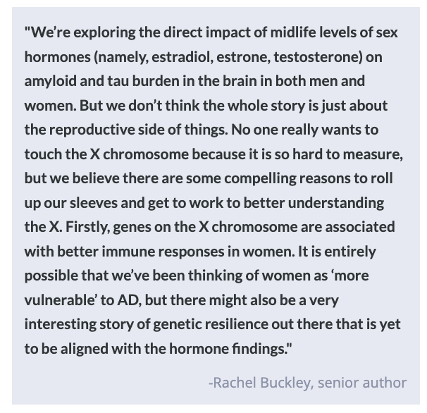
The two hallmarks of Alzheimer's disease (AD) pathology in the brain are the presence of neurofibrillary tangles of tau protein and plaques made of beta-amyloid protein. Research has shown that females have greater levels of tau tangles than males. These differences can be attributed to sex-related risk factors like early onset of menopause and the use of hormone treatment (HT). HT has been suggested as one of the treatments to mitigate cognitive impairment and manage menopausal symptoms like hot flashes, night pain, and urinary issues. However, clinical trials have found evidence suggesting that starting HT after menopause is associated with an increased risk of developing dementia.  

This week in JAMA Neurology, Coughlan and colleagues sought to investigate the relationship between the onset of menopause, age at HT initiation, and Alzheimer’s disease-related pathology in the brain.

How did they do it?

Two hundred ninety-two cognitively unimpaired participants (193 females and 99 males) enrolled in the Wisconsin Registry for Alzheimer's Prevention study were selected for the study. Demographic data (like the age of menopause onset and HT use), lifestyle factors (like education level, menopause severity, and history of hysterectomy), and cognitive performance scores (memory and executive function) were collected. In addition, two specialized Positron Emission Tomography (PET) scans were performed to detect tau tangles and amyloid-beta deposition in parietal, temporal, and occipital brain regions. Linear regression models were run to study the association between sex, age of menopause, and HT use with regional tau and beta-amyloid plaque levels.

What did they find?

Higher levels of cortical tau in parietal and temporal brain regions were found in females than in males, worsened with the presence of beta-amyloid plaques. Precisely in females, younger age at menopause onset, and use of HT post-menopause were associated with high tau deposition. When HT was initiated more than five years after menopause onset, tau levels were significantly higher compared to females who started their HT closer to their menopause age. Finally, cognitive performance was lower in females with late initiation of HT compared to females with earlier initiation of HT. All these results suggest that late initiation of HT can explain the association between HT use and increased levels of tau protein in cognitively unimpaired women.

What's the impact?

Females have an elevated risk of developing dementia due to sex-related factors including the age of menopause and treatment with HT. This study adds to the recommendation of administering HT closer to the menopause onset rather than later. This finding is significant as it can help inform the discussions around hormone-based treatments and AD risk in women. 

Impact of DMT on Brain Function and Connectivity

Post by Lani Cupo

The takeaway

A joint fMRI and EEG experiment reveals the impact of the psychedelic DMT on brain activity, potentially lending insight into the neural correlates of consciousness.

What's the science?

N,N-Dimethyltryptamine (DMT) is a psychedelic that targets serotonin receptors and leads to an altered state of consciousness consisting of vivid imagery without a loss of consciousness. Previous functional magnetic resonance (fMRI) evidence suggests the role of recently-evolved association cortices (transmodal association cortex pole, or TOP) in the effects of DMT. However, these previous findings were obtained with fMRI, which captures not only neural activity but also vascular artifacts and other physiological noise. This week in PNAS, Timmermann and colleagues examined the acute effects of DMT on brain activity with concurrent fMRI and electroencephalograms (EEG), allowing them to correlate methodologies for increased specificity to neural activity. 

How did they do it?

Twenty healthy adults participated in the study, with each participant undergoing two lab visits separated by two weeks. At the first visit, half the participants received a placebo and the other half received DMT, with each participant receiving the other treatment at the second visit. For 30 minutes, fMRI and EEG were acquired simultaneously during rest as the drug or placebo was administered. Participants also provided subjective, real-time ratings of how intense the drug experience was

Initially, the authors examined each modality separately. With the fMRI data, they first calculated static resting state functional connectivity, or in other words they examined the degree to which activity of certain brain regions correlated with activity from other regions across the entire session. For the second fMRI analysis, they used a ‘sliding window’ approach to examine how functional connectivity changed over time throughout the scan, for example calculating correlations for the first minute, then the second minute, etc. They examined how the regional connectivity patterns related to subjective accounts of the intensity of the drug. With the EEG data, the authors looked at a) the signal over the entire period and b) the signal at different times with the sliding window approach. Finally, the authors examined the fMRI and EEG data together by incorporating EEG signals as covariates in regression models of functional connectivity (fMRI). 

What did they find?

With the static functional connectivity approach (fMRI data), the authors found DMT decreased the strength of connections within brain networks as well as the segregation between networks, suggesting regions that comprise a network were less strongly correlated with each other, but more correlated with activity in other regions outside of the network. From the sliding window approach, the authors found subjective reports of greater intensity were associated with increased connectivity among networks. Regions with increased functional connectivity were associated with previously published maps of serotonin receptors, consistent with DMT’s mechanism of action in the brain. 

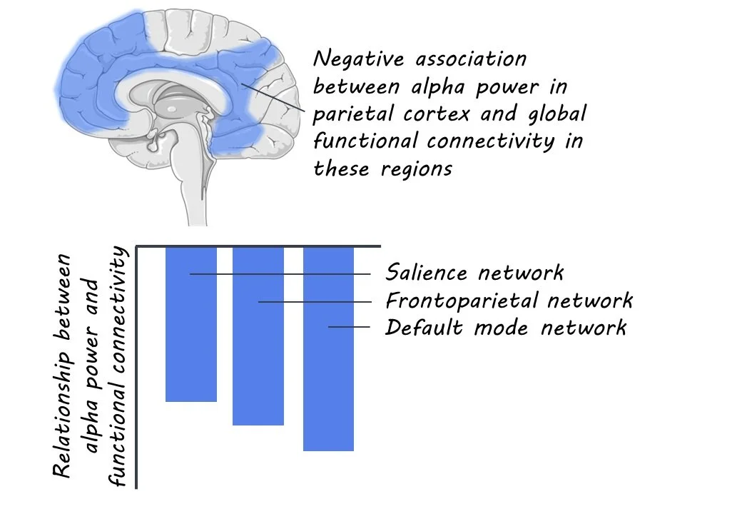
From the EEG data alone, the authors found DMT was associated with decreases in alpha power but increases in gamma and delta power. Alpha waves decrease during sleep, and delta waves are prominent during sleep, suggesting an altered state of consciousness. One hypothesis suggests gamma waves may play a role in communication between neuronal populations, consistent with the decreased segregation of brain networks seen in the fMRI data. More intense subjective ratings were associated with increased delta power but decreased alpha power. Finally, when examining the two modalities together, the authors found that increased delta power was associated with increased functional connectivity among networks across the brain. In contrast, decreased alpha power was related to increased connectivity, including in the TOP, consistent with prior DMT studies. 

What's the impact?

This study confirms previous results suggesting decreased segregation of brain networks while advancing an understanding of how psychedelics alter states of consciousness with multiple modalities. The findings imply that the high-level cortical regions that DMT affects may be necessary for human-specific cognitive traits that underlay human consciousness. 

Access the original scientific publication here