Transgender Listeners Show Reduced Visual Bias When Classifying Voices

Post by Anastasia Sares

The takeaway

While we usually draw on multiple senses and general predictions to inform our perception, this can sometimes backfire, introducing bias into our judgments. This study found that transgender and nonbinary people were less susceptible to visual bias, and better able to classify a person’s vocal range while watching videos of them singing or speaking. This trans advantage could come from more extensive experience with voice-body mismatch in daily life. 

What's the science?

The brain is constantly trying to fuse information from its different senses and make predictions based on that information. Unfortunately, this can sometimes lead to biases when we are only asked to judge based on one sense alone, or if we are confronted with something that doesn’t match our ingrained predictions. One example of this is the McGurk effect, where misleading visual information causes people to perceive a different syllable than the one they heard: a “da” played over audio combined with the visual of someone saying “ba” can result in people reporting that they heard “ga” instead. Visual bias is especially problematic for voice-body mismatches in the context of opera. A person’s body size and shape doesn’t necessarily indicate what range they can comfortably sing, but the stereotypes are strong and can (consciously or subconsciously) influence the roles that opera singers are cast in. This can affect their long-term vocal health and be detrimental to their careers.

Visual biases are not set in stone, however. There is some evidence that they can be mitigated through training, like musicians learning to resist the McGurk effect. One group of people who may have natural sensitivity to voice-body mismatches are the transgender and nonbinary communities, since voice is a strong gender cue and often a source of insecurity or fear of being outed.

Recently in Frontiers in Psychology, Marchand Knight and colleagues showed that, when asked to judge vocal ranges of different speakers, trans and nonbinary people are more resistant to visual biases than their cis peers, making their judgments more accurate.

How did they do it?

The authors conducted an online experiment including a cis group of participants as well as a trans group, which was composed of a mix of trans and nonbinary identities. Participants started by learning about different voice categories used in opera (from low/dark to high/bright: bass, baritone, tenor, alto, mezzo, soprano) and next used this voice-typing scale to rate clips of people speaking and singing. Participants first got the audio-only (no video) versions of the clips, then the visual-only versions (guessing voice type purely based on looks), and finally the full clips with both video and audio. The researchers intentionally chose some actors who they thought might show stronger voice-body mismatches to better measure the effect of visual bias.

What did they find?

Participants were fairly successful at classifying voice type based on hearing the voices in the audio-only condition, but in the visual-only condition they tended to revert to a gender binary (rating videos of female- presenting people around the “mezzo” voice range and male-presenting people around the “baritone” range). The highest and lowest voice types had the most discrepancy between their audio and visual ratings.

When audio and visual were presented together, ratings fell somewhere in between the two previous conditions, showing that the visual information was influencing participants even though they had been asked to rate solely based on the audio. However, the trans participants were better at resisting the visual biases, so their ratings in the audiovisual condition were closer to the audio-only condition. Cis participants’ ratings were pulled more toward the visual information, 30% more so than trans participants. This difference in ability did not seem to be strongly related to demographic differences between the groups or to gender views in general, as far as the researchers could measure. 

What's the impact?

These findings highlight a strength of the trans and nonbinary community, in a time when most research is focusing on the disadvantages they suffer. It also brings up a crucial issue that can affect the vocal health of opera singers, and calls for it to be addressed.

Access the original scientific publication here. 

[Disclosure: The writer of this BrainPost summary is also a collaborator on the publication]

Anxiety is Induced by Activating Microglia, the Immune Cells of the Brain

Post by Rebecca Hill

The takeaway

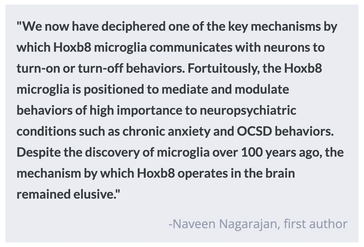
Hoxb8 microglia, the support cells of the brain created by the Hoxb8 gene, play a role in regulating anxiety. When these microglia are activated with light using optogenetics in certain areas of the brain, mice display anxious grooming and freezing behaviors.

What's the science?

Hoxb8 is a gene involved in creating certain microglia, the immune support cells of the brain, but the function of both have yet to be fully elucidated. When the Hoxb8 gene is mutated, or these microglia are removed in mice, they show chronic anxious behaviors and excessive grooming. Recently, in Molecular Psychiatry, Nagarajan and colleagues investigated whether activating these microglia in certain areas of the brain using light has an effect on anxious behaviors in mice.

How did they do it?

To activate the Hoxb8 microglia, the authors used optogenetic stimulation — using light to control the activity of certain cells in the brain. They activated Hoxb8 microglia in specific areas of the brain such as the dorsomedial striatum, the medial prefrontal cortex, the amygdala, and the hippocampus, which have previously been shown to control anxiety in mice. While stimulating these areas of the brain, they measured the behavioral effects; changes in grooming and other anxiety behaviors in different situations. They ran mice through several behavioral tests, measuring the anxiety-behaviors 2 minutes before stimulation, during the 2 minutes of stimulation, then the 2 minutes after stimulation. To measure anxiety levels, they used both a maze and an open field area to test how much time mice would spend in the fear-inducing open areas of the chambers as opposed to comfortable enclosed areas.

What did they find?

Mice groomed themselves when the dorsomedial striatum and the medial prefrontal cortex were stimulated and demonstrated higher levels of anxiety when areas in the amygdala were stimulated. This suggests that grooming is controlled by the former two areas, while anxiety is controlled by the latter area. When the microglia in the hippocampus were stimulated, mice showed both grooming and anxiety behaviors, in addition to increased freezing, which suggests the hippocampus is involved in controlling all three behaviors related to anxiety. Interestingly, when both Hoxb8 microglia and microglia not created by Hoxb8 (non-Hoxb8 microglia) were stimulated at the same time, mice did not display any anxiety behaviors at all. This suggests that Hoxb8 and non-Hoxb8 microglia work together with opposing effects, to control anxiety. Hoxb8 microglia turn off anxiety behaviors (like brakes on a car), and non-Hoxb8 microglia turn these behaviors on (like the accelerator).

In order to reconcile previous findings of anxiety increasing when Hoxb8 microglia are removed, with the current finding that activating Hoxb8 microglia also causes anxiety increase, the authors suggest that optogenetic activation of these Hoxb8 microglia might somehow cancel out their inhibitory effects on anxiety behaviors. While these mechanisms are still not fully understood, they likely involve the neighboring neurons that were activated when the Hoxb8 microglia were stimulated. Either way, these microglia are key in regulating anxiety, potentially in both directions.

What's the impact?

This study is the first to show that Hoxb8 microglia can be used to control anxiety behaviors using optogenetic techniques. It also suggests the reason for having both Hoxb8 and non-Hoxb8 microglia is to finely control anxiety behavior. Anxiety and related mental disorders are widespread both among adolescents and adults, so understanding the way it works within the brain is crucial so that we can better treat chronic anxiety. Studies like these could play a huge part in creating treatments that target these specific microglia and areas of the brain for chronic anxiety disorders.

Associating Heart and Brain Health using MRI

Post by Laura Maile

The takeaway

Evidence suggests a relationship between heart and brain health. The authors identified associations between structural and functional traits of the heart and brain that share genetic signatures with cardiac and brain diseases. 

What's the science?

Cardiovascular disease is often clinically associated with brain diseases, but the underlying genetic, structural, and functional connections between the heart and the brain remain unknown. Magnetic resonance imaging (MRI) has been used to identify structural and functional abnormalities that are associated with disease in individual organs, though few studies have analyzed MRI data from both the heart and the brain to find correlations between the two. 

This week in Science, Zhao and colleagues used multiorgan MRI to examine the connections between heart MRI features and structural and functional patterns in the brain. They then used genome-wide association studies (GWAS) to correlate these findings with genetic variants associated with both heart MRI traits and brain diseases. 

How did they do it?

The authors analyzed MRI data from >40,000 participants in the UK Biobank study. They identified 82 cardiac MRI traits that included measurements of the four cardiac chambers, the ascending and descending aortas, and wall thickness of different regions of the heart. A variety of brain MRI traits were also identified using imaging techniques that examine both structure and functional connectivity of different brain regions at rest and during specific tasks. Next the authors used statistical association and correlation analyses to explore associations between the identified traits. 

The authors then performed GWASs to identify specific genetic variations associated with the cardiac MRI traits. They repeated the GWAS on several different datasets to confirm the associations in a wider population. Next, they completed association and colocalization studies on the significant genetic variants to determine whether the cardiac and brain MRI traits shared genetic signatures. Finally, they sought to determine a genetic causal relationship between the heart and brain by applying Mendelian Randomization to the 82 cardiac MRI traits and several brain-related clinical outcome databases. 

What did they find?

The authors found 4193 significant associations between the 82 identified cardiac MRI traits and brain MRI traits such as cortical thickness, white matter microstructure, and volume of specific brain regions. Associations were also observed between cardiac MRI traits and the functional connectivity between certain brain networks. GWASs identified associations of 49 cardiac MRI traits at 80 genomic loci, which were found to be repeated across several datasets, indicating the generalizability of the findings across populations. They identified genetic variants that were shared across the cardiac and brain MRI traits that had been associated with diseases of the heart and brain. Mendelian randomization analysis revealed a causal relationship between genetic signals associated with heart traits and neuropsychiatric disorders. 

What's the impact?

This study found associations between heart and brain MRI traits that shared common genetic signatures. These findings denote a causal relationship between heart and brain health. This suggests that early intervention and treatment of heart conditions may improve brain health outcomes. 

Access the original scientific publication here