A Novel Subtype of Hypothalamic-Habenula Neurons Drives Aversive Behavior

Post by Trisha Vaidyanathan

The takeaway

The population of glutamatergic excitatory neurons that project from the lateral hypothalamic area (LHA) to the lateral habenula (LHb) is composed of six molecularly, physiologically, and functionally distinct cellular subtypes. One specific subtype, characterized by the expression of estrogen receptor 1 (Esr1+), mediates aversive behavior and a sex-specific maladaptive response to stress.  

What's the science?

The LHA, the LHb, and the prefrontal cortex (PFC) are key nodes in the neural circuit that controls emotional behavior. The LHA is the primary input to the LHb and this LHA-LHb pathway has been shown to send negative signals that mediate avoidance behavior and depression. While recent work has demonstrated that neural populations in other hypothalamic regions are heterogenous, the LHA-LHb cells are still thought to be one homogenous population. This week in Nature Neuroscience, Calvigioni and colleagues used a combination of ex vivo electrophysiology, single-cell RNA sequencing, and mouse genetics to determine if the LHA-LHb population is heterogeneous, characterize any subtypes of LHA-LHb cells, and determine their function in mediating emotional behavior.

How did they do it?

The authors characterized the heterogeneity of LHA-LHb cells using a powerful technique called Patch-Seq, which allowed them to characterize both the electrophysiological properties (via patch clamp recordings) and the gene expression pattern (via single-cell RNA sequencing) of an individual LHA-LHb cell. The authors then used this gene expression data to create Cre mouse lines that enabled them to genetically modify specific LHA-LHb subtypes.

Next, the authors confirmed previous findings that activation of the entire LHA-LHb population leads to aversive behavior using a two-chamber real-time preference test, in which one chamber is associated with optogenetic stimulation and, if aversive, mice will avoid entering that chamber. They next repeated these experiments but only activated specific LHA-LHb subtypes, to identify which subtype is responsible for the aversive behavior. To confirm, they also silenced cells by inhibiting neurotransmitter release via expression of tetanus toxin

Lastly, the authors focused on the cellular subtype marked by expression of estrogen receptor 1 (Esr1+). First, they used high-density electrodes in vivo to compare the PFC response to Esr1+ cell activation with the PFC response to an aversive stimulus in the form of an air puff to the eye. Next, the authors explored the role of Esr1+ cells in a sex-specific maladaptive stress response by exposing male and female mice to an unpredictable foot shock while simultaneously inhibiting Esr1+ cells or, in separate experiments, performing patch clamp electrophysiology to characterize changes in Esr1+ cell properties following the shock stressor.    

What did they find?

First, the authors identified six subtypes of LHA-LHb cells based on the electrophysiological properties obtained with patch-clamp recordings and demonstrated each subtype has a topographical organization across LHA, a unique anatomical projection to LHb, and a unique morphology. Further, single-cell RNA sequencing data revealed unique expression markers for many of these subtypes which the authors then used to generate Cre mouse lines for subtype-specific genetic manipulation.

Next, using optogenetics, the authors demonstrated that only activation of the Esr1+ subtype, but not any other subtype, recapitulated the avoidance behavior caused by activating the entire LHA-LHb population. Further, silencing Esr1+ cells while simultaneously activating the rest of the LHA-LHb population prevented avoidance behavior. This demonstrated that the Esr1+ subtype is necessary and sufficient for mediating the LHA-LHb avoidance behavior. 

Lastly, the authors found that, similar to an external aversive stimulus (air puff to the eye), Esr1+ activation had specific and profound effects on PFC activity, suggesting that Esr1+ cells are a critical component of the broader emotional behavior circuit. The authors also found Esr1+ cells mediate a sex-specific stress response. They demonstrated that unpredictable shocks induced a maladaptive stress response specifically in female mice and this response was reduced if Esr1+ cells were silenced. They also found that unpredictable shocks shifted the intrinsic firing properties of Esr1+ burst-firing cells in female, but not male, mice, suggesting this shift underlies a female-specific susceptibility to stress.

What's the impact?

This study is the first to show that LHA-LHb cells are a heterogenous population and identified six distinct subtypes based on unique physiological, molecular, morphological, and anatomical markers. Further, they demonstrate that a specific subtype of LHA-LHb cells marked by Esr1 expression is necessary and sufficient for aversive behavior and sex-specific stress responses. Broadly, this research reveals the importance of characterizing the diversity of neuron subtypes that underlie complex emotional behaviors. 

Access the original scientific publication here

Transcriptomic Changes in Cellular Communities In the Brain Contributes to Alzheimer’s Disease

Post by Soumilee Chaudhuri

The takeaway

Major brain cell types — neurons, oligodendrocytes, endothelial cells, etc. — individually and synergistically contribute towards molecular changes seen in the aging human brain in Alzheimer’s Disease (AD). A high-resolution transcriptomic map in the aging human brain unraveled 1) diverse cell populations associated with AD and 2) how networks of cellular communities coordinate to alter biological pathways, ultimately leading to AD.

What's the science?

Alzheimer’s Disease is an irreversible, neurodegenerative illness, with a complex pathophysiology. Amongst many unknowns in the molecular mechanisms of AD, is the specific contribution of major cell types in the aging brain and how this might trigger AD-related dementia. Even so, knowledge of the contribution of different brain cells in AD pathogenesis has advanced over the last decade due to the advent of high-resolution technologies such as single-cell RNA sequencing. We know that perturbation in transcriptomes (i.e., full range of mRNA molecules produced) of major brain cell types and subtypes like excitatory and inhibitory neurons, oligodendrocytes or microglia for example, synergize to cause events that lead to molecular changes seen in AD. However, we do not know the specific contributions of each cell subtype to this disease due to limited sample size and a lack of robust and sensitive technologies powerful enough to capture interindividual as well as cell-specific diversity in AD brain. This week in Nature Neuroscience, Dr. Cain and colleagues unravel the distinct cellular architecture of the aging brain prefrontal cortex in AD as well as how cells interact, using combined bulk and single cell RNA sequencing analyses and novel bioinformatics pipelines.

How did they do it?

The authors used a powerful approach to get insights about coordinated multicellular communities in the AD brain. They used a) integrative transcriptomics (bulk and single-cell RNA sequencing) as well as b) a robust analytical approach (CellMod: a deconvolution tool that allows estimation and projection of single cellular landscapes from a limited set of individuals to a higher number of individuals). The authors used single-cell data from 24 individuals from the ROSMAP cohort to generate a cellular map of the aging Dorsolateral Prefrontal Cortex (DLPFC) brain region, and used that as input to their novel CellMod deconvolution algorithm to estimate cellular compositions in an independent set of 638 individuals with bulk RNAseq data. Then, network analysis within this model revealed cellular sets and subsets and interacting cellular communities across individuals with AD vs. controls; and advanced statistical modeling associated known AD traits and risk factors to these identified cell subsets and communities across the case (AD) vs control (no AD) groups.

What did they find?

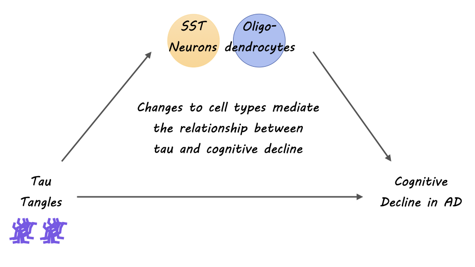
The authors identified specific subpopulations of cells associated with AD pathophysiology. Primarily, the authors discovered oligodendrocyte transcriptional pathways and a downregulation in somatostatin-producing neurons (SST neurons) as perturbed in AD pathogenesis. From the innovative cellular map of the neocortex that the authors constructed, they found interesting insights about oligodendrocyte diversity and interactions, implicating oligodendrocytes  —the myelinating cells of the brain — as a strong cellular contributor to AD. Further, they identified that oligodendrocyte expression from both positive (Oli.4) and negative oligodendrocyte (Oli.1) cells was strongly associated with tau pathology and cognitive decline in AD. Multiple shared pathways within cellular communities were identified and related to known risk factors of AD, as a way of validating that AD indeed, is a pathophysiologic process with multiple interacting cell types. Overall, the findings of this study pinpoint the importance of approaching our understanding of AD through a lens of interacting multicellular communities and networks. 

What's the impact?

This study is the first to show that cell specific as well as coordinated cellular and sub-cellular interactions in the aging brain may contribute to a diseased microenvironment in AD. Additionally, the authors find evidence of the contribution of communities of oligodendrocytes to cognitive decline and tau burden in Alzheimer’s Disease. The result of this study extends research on cellular and subcellular heterogeneity in the diseased aging brain and helps to inform therapeutic targets for AD and dementia.

Access the original scientific publication here

Understanding How Decisions Are Terminated

Post by Kulpreet Cheema

The takeaway

Little is known about how decisions are terminated and translated into actions or plans. This study provides evidence that the superior colliculus (SC), a midbrain structure involved in eye movements and orienting behaviors, plays a crucial role in terminating decisions.  

What's the science?

Previous research has shed light on how the brain accumulates evidence before reaching a decision. This process can be modeled as a stochastic drift-diffusion process or bounded random walk. Neurons in the lateral intraparietal area (LIP) have been shown to accumulate noisy evidence during decisions. However, how this process is terminated and translated into a specific action is still unknown. The SC is known for its role in generating eye movements, and is directly coupled to the LIP; the LIP projects to the SC and the SC projects back to LIP via the thalamus. In a study published in Neuron, researchers investigated the role of SC in applying a decision threshold to the accumulation of evidence represented in the LIP.

How did they do it?

The researchers recorded simultaneous neural activity in the LIP and SC while monkeys performed a motion-discrimination task. In the motion discrimination task, the monkeys were trained to make eye movements based on the direction of a moving stimulus. Researchers used high-density multi-channel electrodes to capture the firing rates of functionally similar neurons in both areas during the decision process. To directly test the involvement of the SC in decision termination, researchers performed focal inactivation by temporarily inactivating the SC using small muscimol injections. They analyzed behavioral measures and neural recordings from the LIP during SC inactivation.

What did they find?

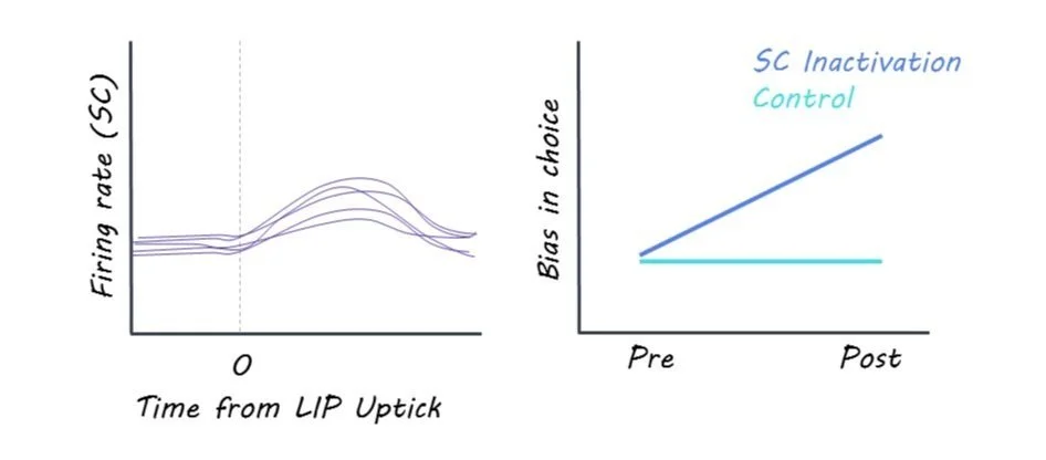
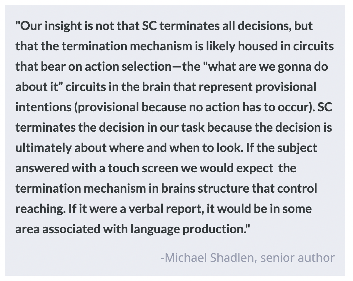
Researchers found evidence that the LIP and SC exhibited different dynamics during decision-making, and that the SC implements the decision threshold. The researchers observed that bursts of activity in the SC terminated the decision process, as opposed to the accumulation signals in LIP. The bursts in the SC were triggered by upticks in excitatory input and were associated with the termination of the decision. When the SC was inactivated, the termination mechanism was impaired, leading to slower, biased decisions and prolonged evidence accumulation in LIP. These findings suggest that, while evidence may accumulate in the LIP during decision-making, decision termination occurs in an area responsible for action selection; in this case, the SC, as the decision is about moving the eyes.

What's the impact?

This study sheds light on how the brain makes decisions and transforms evidence into actions. Understanding how decisions are terminated is crucial for comprehending the entire decision-making process. By identifying the role of the SC in decision termination, the study highlights the importance of a region known for action selection in decision termination. The findings have implications for understanding decision-making processes in humans and other primates.