Different Patterns of Gene Mutation in Neurons and Glia During Aging

Post by Lani Cupo

The takeaway

Neurons and glia show different patterns of gene mutations during the aging process in humans, suggesting different processes may underlie age-related genetic mutations in different cell types. 

What's the science?

The underlying cellular and molecular mechanisms contributing to the process of typical aging (in the absence of disease) are still poorly understood. While aging-related genetic mutations in neurons have been investigated, glia, responsible for everything from providing brain structure to maintaining homeostasis, represent more than half of the cellular content of the brain and have yet to be examined for gene mutations in the aging process. This week in Cell, Ganz and colleagues present a characterization of mutations in neurons and oligodendrocytes (glia that produce myelin in the brain), finding different patterns of mutations between the two cell types.

How did they do it?

The authors extracted oligodendrocytes from the prefrontal cortex of 13 deceased individuals aged 0.4 - 83 years and accessed genome data from the neurons of 19 individuals (12 of whom overlapped with their oligodendrocyte donors). They used a method called single-cell whole-genome sequencing to identify the genetic code of these cells individually. Then, they used an algorithm to automatically identify two types of mutations in the genomes of the cells: single-cell whole-genome sequencing (sSNVs) and small insertions and deletions (indels). sSNVs occur when a single nucleotide is switched for another, and are acquired over the lifespan, rather than being inherited. Indels refer to insertions or deletions of nucleotides in the genome. With these data, the authors first estimated annual rates of accumulation of both sSNVs and indels. They also examined where in the genome the mutations occurred. Finally, they compared the patterns of mutations they observed with genomes from a database of tumor cells.

What did they find?

The authors found that oligodendrocytes exhibited higher rates of sSNV accumulation, but lower rates of indel accumulation compared to neurons. Deletions of nucleotide base pairs were more common than insertions in both cell types, but there were also differences in the kinds of deletions between the cell types. Oligodendrocytes mostly showed deletions of a single base pair whereas neurons showed deletions of 2-4 base pairs or 1 base pair insertion compared to the glial cells. At birth there was no significant difference between sSNVs or indels between cell types, suggesting that the changes were acquired over the lifespan and relevant to aging. The fact that profiles of mutations differed between cell types provided evidence that different mechanisms underlie aging between cell types. 

The authors also found opposite patterns in the placement of mutations between cell types. Mutations in oligodendrocytes were found to be more prevalent in intergenic regions, or parts of the genetic code that are not transcribed into proteins. In contrast, mutations in neurons were found to be more prevalent in regions in genes - regions that are transcribed into proteins. As a result of the placement, mutations in neurons were also found to have more of a functional effect on the cell. When comparing the mutation profiles of oligodendrocytes and neurons to cells from tumors, the authors found that densities of sSNVs in oligodendrocytes correlated with those in all cancer types, but neuronal mutations did not correlate with mutations in cancer cells, suggesting the mechanism contributing to mutations in glia cells, but not neurons, may be related to tumor formation.

What's the impact?

For the first time, this study compares genetic mutations in oligodendrocytes and neurons throughout the typical aging process. They found differences in the type and placement of mutations between cell types, suggesting different mechanisms contribute to the mutations. In time this may help us identify the specific mechanisms underlying what goes wrong in aging and neurodegenerative diseases.

 Access the original scientific publication here.

A Mobile Messaging Intervention Can Improve Depression Symptoms in Older Adults

Post by Lila Metko

The takeaway

Adequate mental health care for older adults is a growing concern in low and middle-income countries. A new mobile messaging intervention, developed to combat this issue, was able to help older adults with depressive symptoms in Brazil by improving their condition to a less depressed state.

What’s the science?

Given that 69% of the world's older population live in low and middle-income countries, and that the healthcare workers in these countries are already overstretched with a heavy workload, a virtual yet efficacious option for mental health treatment is needed. This week in Nature Medicine, Scazufca and colleagues assess the effectiveness of a mobile messaging intervention in older adults with depressive symptoms, that uses two techniques called psychoeducation and behavioral activation. Psychoeducation is a specific way of educating patients about their condition, which can have a therapeutic value to them. Behavioral activation is the encouragement of the individual by their mental health provider to engage in a constructive activity like going for a walk in nature or calling a good friend. 

How did they do it?

The researchers recruited 603 participants who were over the age of 60, experienced depressive symptoms, and had access to WhatsApp mobile messaging. These participants were recruited from underprivileged areas in a major metropolitan region of Sao Paulo, Brazil. Participants were randomly separated into two groups, an intervention arm, and a control arm. The intervention arm participants received a self-help mobile-based intervention developed based on the principles of psychoeducation and behavioral activation. Forty-eight WhatsApp messages in either video or audio format (depending on the message) were sent to the participants so that they received a message twice a day, four days a week for six weeks. The audio messages were 3 minutes long, and contained fictional characters who discussed symptoms of depression and potential interventions, mentioning fictional situations of interventions that had been beneficial in the past. The visual messages were summary diagrams designed to reinforce the audio message content. The control group received only one six-minute message that briefly discussed symptoms of depression and how to manage them.

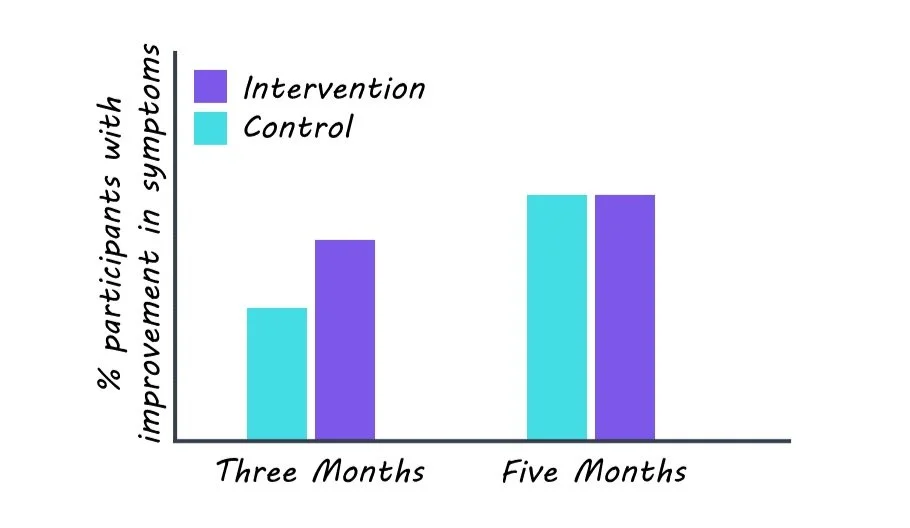
Participants’ level of depressive symptomology was measured at baseline, three months after the intervention, and five months after the intervention. The two measures assessed related to symptomology were: improvement in depressive symptomology and reduction in depressive symptomology. Improvement in depressive symptomology constituted having a score on the depressive symptom questionnaire below a specific value and reduction in depressive symptomology constituted having a reduction in their depressive symptomology questionnaire score by at least 50%.   

What did they find?

Participants in the intervention arm had improvement and reduction in depressive symptomology at three months but not at five months. A complier average causal effect (CACE) analysis indicated that, for the three-month time period, as the number of messages opened by the participants increased (compliance), so did the effect of the intervention.

What’s the impact?  

Many low and middle-income countries such as Brazil have a treatment gap for mental health. This is especially concerning due to the growing population of older adults, and the need for mental health care. Roughly 30% of Brazilian older adults in low-resource settings experienced symptoms that could indicate clinical depression. This research indicates that self-help interventions for depression are feasible and efficacious in low and middle-income populations.

Access the original scientific publication here.

Risk Factors Affecting Brain Regions Vulnerable to Aging and Disease

Post by Meagan Marks

The takeaway

The “LIFO” network is a collection of high-functioning brain regions most susceptible to decline by aging and disease. Particular genes and modifiable risk factors such as diabetes, pollution, and alcohol intake are shown to be linked to the vulnerability of these regions.

What's the science?

Regions of the brain associated with high-process functions such as memory, attention, and execution tend to degenerate earlier and faster than the rest of the brain. These regions are also the last to develop during adolescence, coining them the “last in, first out” (LIFO) network. It is known that these LIFO regions are especially vulnerable to diseases like Alzheimer’s, Parkinson’s, and Schizophrenia, however, the genetic and modifiable risk factors that influence the sensitivity of these regions remain unknown. This week in Nature Communications, Manuello and colleagues aimed to identify these factors via statistical analysis to understand further how the LIFO network is regulated and to determine potential behaviors that may shield or exacerbate its decline.

How did they do it?

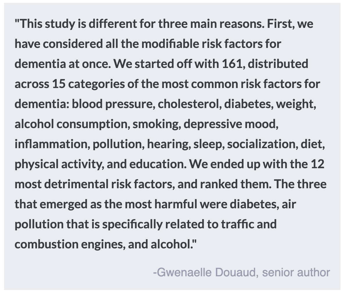
To determine the specific genes and modifiable risk factors linked to the LIFO network, the authors ran a series of statistical tests using data from the UK Biobank, a large-scale biomedical database that holds genetic, lifestyle, and health information of thousands of participants. To begin, the authors first calculated the grey matter volume of the LIFO regions of nearly 40,000 participants using brain scans obtained from the biobank. This allowed the authors to determine how much degeneration had taken place (less volume = more degeneration). Next, using computational analysis, the authors were able to sift through the genomes of participants to identify which genes were significantly associated with the grey matter volume of the LIFO network and which variants, or versions, of these genes were significantly associated with a lower volume. Finally, the authors looked at the association between the LIFO network and 15 modifiable risk factor categories that were previously linked to dementia. The authors were able to use the health and lifestyle data of biobank participants to do so, statistically determining which of these factors were significantly associated with LIFO network grey matter volume.

What did they find?

After genomic analysis, the authors identified seven gene clusters, or groups of genes, that were linked to the LIFO network. The top gene variants in these clusters (3,934 total) were from genes that regulate immune cell trafficking, inflammation, and neurogenesis, genes that have been linked to blood pressure, sleep duration, and cognitive performance, and genes located in a genetic region associated with Alzheimer’s disease and other neurodegenerative disorders. In total, these results suggest that individuals with specific variants of the genes identified may have a LIFO network more vulnerable to disease and aging.

Regarding modifiable risk factors, the authors found that 12 of the 15 modifiable risk factor categories had at least one factor that significantly affected the LIFO brain network. Taken together, these 12 factors significantly explained 1.5% of the vulnerability of the network after age and sex were removed. Of these factors, the authors determined that diabetes, alcohol intake, and pollution by nitrogen dioxide were the most harmful to the LIFO regions. This suggests that individuals who have been diagnosed with diabetes, who consume copious amounts of alcohol, or who have high exposure to nitrogen dioxide pollution may be at a higher risk of cognitive degeneration in these brain regions. 

What's the impact?

This study is the first to identify specific genetic and modifiable risk factors associated with the LIFO brain network, which contains the high-functioning regions most vulnerable to decline by aging and disease. Determining the specific gene variants and modifiable risk factors that are associated with LIFO regions may help to identify individuals at higher risk for cognitive decline. Recognizing these factors will allow patients and providers more time to protect against potential decline and help explain the biological mechanisms behind degeneration.