The Placebo Effect is Driven by Brain Regions Associated with Value and Motivation

 Post by Lila Metko 

The takeaway 

Placebo effects are forged by internal neural processes that bring about improved health. Interestingly, those who experience a placebo effect do not show decreased activity in brain regions associated with early pain perception, but rather brain systems relating to value and motivation. 

What's the science?

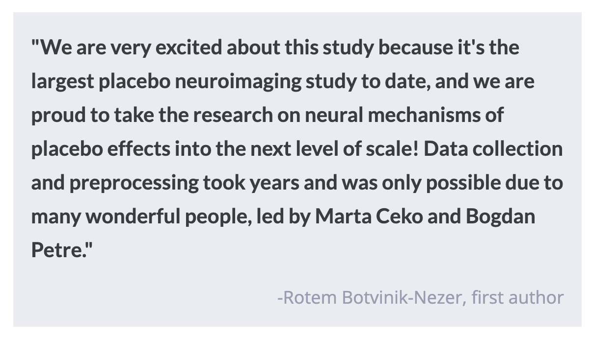
Many current theories posit that pain is primarily a top-down phenomenon. Higher-level brain regions integrate contextual and affective information with sensory signals about potentially harmful stimuli to create predictions about future pain. The placebo effect is when someone’s physical or mental health improves after taking a placebo or 'dummy' treatment. Previous brain imaging studies have shown reductions in neural activity in pain processing regions such as the medial and ventrolateral thalamus and dorsal posterior insula to be correlated with analgesia after the administration of a placebo (the reduction of a pain sensation). This week in Nature Communications, Botnivik-Nezer and colleagues tracked fMRI activity across two different pain signatures (i.e., patterns of brain activity) in the brain in placebo and control conditions to understand the brain mechanisms involved in the placebo effect.

How did they do it?

Approximately 395 participants received an application of a control cream with “no effects” or a cream presented to them as a pain-relieving drug (placebo) on different fingers of the left hand. Conditioning paradigms were then used to strengthen the placebo effect. To illustrate, in one paradigm participants were exposed to a range of thermal stimuli on both the control and placebo fingers, but unbeknownst to the participant, the stimuli were not as strong on the placebo finger. Participants then had the creams reapplied such that four fingers had a cream: two placebo, and two control, so that the authors could test for mechanical pain and thermal pain in both control and placebo condition fingers. They were then monitored in an MRI scanner a test task, in which they would be presented with a stimulus and would need to respond with a rating of its intensity and degree of unpleasantness. The stimuli were presented at low, medium, and high intensities with a randomized order of which pain modality was being examined (thermal or mechanical pain). The authors then calculated the extent of placebo analgesia for each participant and compared it to changes in brain activity in two fMRI brain signatures for pain, the Neurologic Pain Signature (NPS) and the Stimulus Intensity Independent Pain Signature (SIIPS). The NPS is a signature more associated with the initial, unprocessed perception of painful stimuli based on the sensory input, whereas the SIIPS is more associated with neural structures that provide the top-down, internal evaluation of pain. Additionally, the authors compared the placebo effects on a conditioned pain modality (thermal) to an unconditioned pain modality (mechanical) to test for transfer of the placebo effect to unreinforced modalities. 

What did they find?

Participants experienced a placebo effect and reported less pain in the placebo compared to the control condition. Focusing on the neural response, the authors found that while the NPS fMRI score increased during both thermal and mechanical stimuli and higher NPS score correlated with increased stimulus intensity, there was no effect of placebo analgesia on the NPS score for either modality. Interestingly, in both thermal and mechanical modalities the placebo scores were significantly lower for the SIIPS neural signature than the control scores. This indicates that the placebo effect is likely mediated by higher-level pain processing systems, which are represented in the SIIPS but not in the NPS. Additionally, it was found that participants’ ratings for mechanical pain, a modality that had not been conditioned with the placebo cream, were lower in the placebo finger than the control. When the average placebo effect for participants was compared between the two modalities, there was no difference between the thermal and mechanical placebo effect scores, providing further evidence that the placebo effect is transferable across pain modalities. However, a correlation between expected results for the placebo cream (pre-testing) and test ratings was only found in the thermal (conditioned) modality. 

What's the impact?

This is the first study to investigate the effect of placebo analgesia on brain activity using a large sample size and two well-validated brain signatures for pain. The findings of this study suggest that the placebo effect is driven by higher-order brain systems that integrate value into pain perception rather than lower-level systems that are active earlier in pain processing. This is important because, with more detailed knowledge about the placebo mechanism, practitioners may be able to focus treatments on higher-level cognitive processes to treat pain.  

Neuromodulatory Control of Bionic Limbs Enables Natural Walking Patterns

Post by Shireen Parimoo

The takeaway

Prosthetic or bionic limbs have traditionally required pre-programmed gait controllers to enable movement following limb amputation, but their use has not quite resembled natural walking gait. Now, researchers have shown that biomimetic gait (i.e., a gait that resembles that of biologically intact limbs) can be restored by providing neuromodulatory input to bionic limb controllers in real time through an innovative surgical procedure. 

What's the science?

Bionic limbs have been around in some form for thousands of years and have undergone incredible advances in recent decades. Today, bionic limbs use controllers pre-programmed with gait algorithms to assist individuals with locomotion. However, they do not use sensory neural inputs to adjust gait, nor do they provide enough information about the position and velocity of the bionic limb to the user, often causing them to rely on visual inputs to control their movement. As a result, bionic limbs have not been able to fully mimic the natural gait of biologically intact limbs.

One reason why neurofeedback has not been fully integrated into bionic limb function is that the peripheral tissue that’s important for neuromuscular control is removed during the amputation surgery. The agonist-antagonist myoneural interface (AMI) is a procedure that uses the remaining biological tissue by connecting agonist (shortening) and antagonist (lengthening) muscles in the amputated region, to replicate the interaction between agonist and antagonist muscles present in biologically intact limbs. This week in Nature Medicine, Song and colleagues used AMIs to achieve biomimetic gait in individuals with bionic limbs without relying on pre-programmed gait controllers.

How did they do it?

Participants underwent the below-knee AMI amputation or a non-AMI amputation (i.e., control group). During the AMI surgery, the lateral gastrocnemius and tibialis anterior were connected to facilitate ankle control while the peroneus longus and tibialis posterior were linked to enable foot rotation. The AMI participants were fitted with a bionic ankle with a controller that monitored the angular position and speed of the ankle and an electromyography sensor that provided neurofeedback to the controller. Inputs from both the sensor and the bionic limb were processed by the controller for bidirectional feedback.  

The authors measured walking speed and gait on a flat surface, on a slope (incline and decline), up and down stairs, and over small obstacles on the ground. Gait was assessed by examining the angular position and velocity (i.e., state) of the bionic ankle, while the torque angle (twisting or rotating) of the bionic ankle was used to quantify the degree of biomimetic gait. The ankle state and torque angle were used to calculate the amount of power and net work (total force) generated by the bionic ankle across different terrains. These measures were compared between the AMI and control groups, as well as with publicly available data on the gait patterns of individuals with biologically intact limbs.

What did they find?

The AMI group walked faster and showed a higher degree of biomimetic gait than the control group. For example, ankle power tends to increase with walking speed in biologically intact limbs, and this increase was seen only in the AMI group. The maximum walking speed of participants in the AMI group was also on par with that of individuals with biologically intact limbs. When participants were divided according to the magnitude of the agonist-antagonist muscle afferents, those in the control group had little to no residual muscle afferents while those in the AMI group had ‘moderate to high’ levels of afferents. Conversely, the torque angle trajectory of control participants resembled stiff ankles and non-biomimetic gait, while the AMI group were closer to exhibiting natural walking dynamics. Thus, using the residual muscle afferents to provide neurofeedback to bionic limbs enables biomimetic gait.

The AMI group adapted to different terrains faster than the control group. Their ankle peak power and net work increased while walking up an incline, while their shock absorption increased while descending stairs. The control group, on the other hand, did not show these adaptations and had a more limited range of motion. In the presence of obstacles, the AMI group increased dorsiflexion during the upswing of the ankle, followed by increasing propulsive power and net work on the recovery step, which allowed them to better maintain their walking speed. Dorsiflexion did not increase in the control group (and even decreased for some participants) in the presence of obstacles, which in turn slowed them down. Together, these results indicate that the neuromodulatory mechanism in the AMI amputation allowed participants to successfully adapt to different types of terrain and environmental obstructions.

What's the impact?

This study is the first to use AMI to demonstrate biomimetic gait and natural walking patterns in a bionic limb that relies entirely on neuromodulatory mechanisms. These findings show promise for AMI amputation to change the landscape of neuroprosthetic limb design and push the field closer to fully restoring biomimetic motor functioning following surgical amputation.

Access the original scientific publication here.

How Stress Impacts Learning and Memory – The Good, the Bad, and the Biological

Post by Rebecca Hill 

The takeaway

Stress, in particular chronic stress, is often linked to poor outcomes for learning and memory. However, depending on the situation, the right amount of stress may have positive learning outcomes.

What does stress do to the brain and body?

We all experience stress to some degree in our everyday lives. Stress impacts us mentally and physically with profound effects throughout the entire body. First, the stressor activates brain regions such as the hypothalamus and pituitary, which activates the sympathetic nervous system – commonly known as the fight-or-flight response. This causes the release of adrenaline, leading to increased attention and better memory storage. This helps you remember the most important information about a stressor so you can cope with similar future events. Typically, this is a useful response, but if the stress is too intense, it can lead to stress and anxiety disorders, such as post-traumatic stress disorder (PTSD).

How is learning impacted by stress?

Learning and memory can be positively or negatively impacted by stress, depending on the circumstances. If you’ve ever experienced a strong negative event, you can likely remember extremely specific details about where you were and what you were doing at the time of that event. This is due to the sympathetic nervous system’s incredible ability to help you form detailed memories of stressful events. However, you may also have times in your life when you were so stressed out, you hardly remember them. This usually happens when stress is chronic or does not overlap with the timing of the information to be learned. In this way, stress can impair an individual’s ability to form memories. Using functional magnetic resonance imaging (fMRI), some researchers found that in these cases, stress can even reduce brain activity in the regions associated with memory, such as the hippocampus.

When does stress lead to poor outcomes?

Many current studies focus on stressful situations that can worsen learning capabilities. A research study found that depressed students dealing with stress have worse memory and find it more difficult to learn during school. One study used a virtual reality simulation to see how stress impacts individuals’ abilities to complete a navigational planning task. They found that participants who were stressed had more disrupted memory than control participants. Another study found that acute stress particularly disrupts spatial learning and memory. Stress can have even worse impacts on the developing memories of children. For example, excessive screen time can cause chronic stress which leads to increased anxiety and negatively affects learning and memory. Both long-term and short-term stress can lead to negative impacts, both early and later on in life.

Could stress have positive outcomes?

While stress is often considered negative, a recent paper discussed the possibility of stress having a positive influence on learning and memory. For example, completing a challenging task initially causes a small amount of stress, but can lead to learning after overcoming the challenge. The researchers emphasized that the key to stress leading to positive outcomes is both the context of the stress and how an individual interprets the stressor – a calm, quiet environment is much more likely to lead to a positive learning outcome than a noisy one. Also, when an individual approaches a stressful situation with a growth-oriented mindset, rather than one that expects a negative outcome initially, it can help the individual cope with stress. So, while stressful situations are often unavoidable, they can be used as opportunities to learn and grow, especially if the stress is moderate, in the right context, and approached with a positive mindset.

References +

Aprilia, A., & Aminatun, D. (2022). INVESTIGATING MEMORY LOSS: HOW DEPRESSION AFFECTS STUDENTS’ MEMORY ENDURANCE. Journal of English Language Teaching and Learning, 3(1), 1–11. https://doi.org/10.33365/jeltl.v3i1.1719

Brown, T. I., Gagnon, S. A., & Wagner, A. D. (2020). Stress Disrupts Human Hippocampal-Prefrontal Function during Prospective Spatial Navigation and Hinders Flexible Behavior. Current Biology, 30(10), 1821-1833.e8. https://doi.org/10.1016/j.cub.2020.03.006

Meyer, T., Quaedflieg, C. W. E. M., Bisby, J. A., & Smeets, T. (2020). Acute stress – but not aversive scene content – impairs spatial configuration learning. Cognition and Emotion, 34(2), 201–216. https://doi.org/10.1080/02699931.2019.1604320

Neophytou, E., Manwell, L. A., & Eikelboom, R. (2021). Effects of Excessive Screen Time on Neurodevelopment, Learning, Memory, Mental Health, and Neurodegeneration: A Scoping Review. International Journal of Mental Health and Addiction, 19(3), 724–744. https://doi.org/10.1007/s11469-019-00182-2

Rudland, J. R., Golding, C., & Wilkinson, T. J. (2020). The stress paradox: How stress can be good for learning. Medical Education, 54(1), 40–45. https://doi.org/10.1111/medu.13830

Schwabe, L., Hermans, E. J., Joëls, M., & Roozendaal, B. (2022). Mechanisms of memory under stress. Neuron, 110(9), 1450–1467. https://doi.org/10.1016/j.neuron.2022.02.020