Oxytocin and the Social Brain

Post by Lila Metko

The takeaway

Oxytocin has long been known as a peptide involved in promoting social interactions between individuals, in animals and humans alike. Recently, studies have pointed to the conclusion that oxytocin may not be as involved in human social interaction as was previously believed.

What is oxytocin?

Oxytocin is a neuropeptide that acts in both the central and peripheral nervous system. Before it was known for its role in social cognition, it was primarily known as a homeostatic hormone with some effects on sexual behavior. A 1991 study by Popik and Vetulani changed this view when they showed that antagonists of oxytocin receptors improved social memory while the delivery of oxytocin impaired it. Now, oxytocin is known as a major regulator of the social brain. However, the extent to which oxytocin affects different types of social interactions is not well understood. In this topic overview we explore the current literature about oxytocin and social behaviors.

How does oxytocin impact the parental bond?

Oxytocin has a strong impact on the bond between a parent and their offspring. Human mothers suffering from postpartum depression who were given oxytocin intranasally have shown greater protective responses towards their newborns than before oxytocin administration. Additionally, mothers with irregularities in the gene that codes for the oxytocin receptor have exhibited less sensitive parenting behaviors toward their children. Perhaps the most convincing piece of evidence that oxytocin plays a role in this bond is work showing that oxytocin triggers maternal behavior in rats that had never experienced a pregnancy. Interestingly, this could only occur when the rats were primed with estrogen, and the oxytocin dose was extremely high. However, a 2010 paper by MacBeth and colleagues revealed a surprising twist: mice lacking oxytocin receptors still exhibited typical maternal behavior. Further investigation by the same research group uncovered a more nuanced role for oxytocin: while these mice were indeed capable of displaying typical maternal behaviors, oxytocin's primary function appeared to be lowering the threshold at which maternal behaviors were initiated. More recent work (2022) has added another layer of complexity, demonstrating that maternal behaviors in these oxytocin receptor-deficient mice were specifically diminished after exposure to stressful environments. These findings collectively suggest that while oxytocin plays a significant role in maternal behavior, its influence is part of a more intricate and multifaceted system that regulates parental care in mammals.

How does oxytocin affect bonding in other relationships?

If the conclusions are mixed about oxytocin and the parental bond, what about oxytocin and other social contexts? Research in prairie voles, which form long-lasting partnerships with their mates, has shown that oxytocin infusions can bring about a partner preference even in the absence of mating, and that oxytocin antagonists can abolish partner preference in a mated pair. A human study by Kosfeld and colleagues showed that intranasal oxytocin was able to increase trust in a randomly selected ‘trust game’ partner. However, recently it was found that attempts to replicate the Kosfeld study were unsuccessful. In conclusion, while oxytocin may establish partner preference in the highly social species of prairie voles, it has not yet been shown to translate to human concepts like trust.

Oxytocin can also impact social cognition — mental actions such as thinking, learning about, or understanding a social interaction. Mice genetically lacking oxytocin have no social memory (could not distinguish familiar and novel mice) but when given oxytocin, social memory was restored. It was later shown that this action occurred through receptors in the amygdala, a brain region involved in emotional memory. Some research in humans suggests that there are sex differences in the effects of oxytocin on social cognition and behavior and that these effects are context-dependent. For example, evidence suggests that the 'prosocial' effects of oxytocin are more prominent in males.

How does oxytocin affect mood?

Interestingly, oxytocin can affect more than just social behaviors, but also social-related phenomena like emotions. In a study of 37 healthy men put into a stressful situation (a mock job interview), it was shown that intranasal oxytocin had anxiety-relieving effects. In a mouse model of hormone-induced postpartum depression, it was found that oxytocin receptor levels were low in certain brain regions. Additionally, intranasal oxytocin can affect the mood of women with postpartum depression, although how it affects mood is unclear. Some studies show that oxytocin improves mood in postpartum women, while another study showed that oxytocin exacerbated depressive symptoms.

What can we conclude from these findings?

In the current literature on oxytocin’s impact on the social brain, there is a large amount of variation in outcomes. However, it is clear that oxytocin affects social pathways in the brains of both humans and animals. The extent to which these animal studies can translate to humans needs to be explored further.

We are undeniably a social society, and social interactions make up a large part of many people’s daily lives. Additionally, the symptom profiles of multiple mental health disorders, such as schizophrenia and autism spectrum disorder, include deficits in social cognition. Therefore, from a treatment-focused perspective, understanding the role of oxytocin and its impact on social behavior is crucial.

References +

Leng, G., Leng, R. I., & Ludwig, M. (2022). Oxytocin-a social peptide? Deconstructing the evidence. Philosophical transactions of the Royal Society of London. Series B, Biological sciences, 377(1858), 20210055. https://doi.org/10.1098/rstb.2021.0055

Popik, P., & Vetulani, J. (1991). Opposite action of oxytocin and its peptide antagonists on social memory in rats. Neuropeptides, 18(1), 23–27. https://doi.org/10.1016/0143-4179(91)90159-g

Zhu, J., Jin, J., & Tang, J. (2023). Oxytocin and Women Postpartum Depression: A Systematic Review of Randomized Controlled Trials. Neuropsychiatric disease and treatment, 19, 939–947. https://doi.org/10.2147/NDT.S393499

Bakermans-Kranenburg, M. J., & van Ijzendoorn, M. H. (2008). Oxytocin receptor (OXTR) and serotonin transporter (5-HTT) genes associated with observed parenting. Social cognitive and affective neuroscience, 3(2), 128–134. https://doi.org/10.1093/scan/nsn004

Macbeth, A. H., Stepp, J. E., Lee, H.-J., Young, W. S. III, & Caldwell, H. K. (2010). Normal maternal behavior, but increased pup mortality, in conditional oxytocin receptor knockout females. Behavioral Neuroscience, 124(5), 677–685. https://doi.org/10.1037/a0020799

Rich, M. E., deCardenas, E. J., Lee, H., & Caldwell, H. K. (2014). Impairments in the Initiation of Maternal Behavior in Oxytocin Receptor Knockout Mice. Plus ONE. 9(6), e99839. https://doi:10.1371/journal.pone.0098839

Tsuneoka, Y., Yoshihara, C., Ohnishi, R., Yoshida, S., Miyazawa, E., Yamada, M., Horiguchi, K., Young, W.S., Nishimori, K., Kato, T., Kuroda, K.O. (2022). Oxytocin Facilitates Allomaternal Behavior under Stress in Laboratory Mice. eNeuro, 9(1), 0405-21. https://doi: 10.1523/ENEURO.0405-21.2022

Kosfeld, M., Heinrichs, M., Zak, P. J., Fischbacher, U., & Fehr, E. (2005). Oxytocin increases trust in humans. Nature, 435 (7042), 673–676. https://doi.org/10.1038/nature03701

Heinrichs, M., Baumgartner, T., Kirschbaum, C., & Ehlert, U. (2003). Social support and oxytocin interact to suppress cortisol and subjective responses to psychosocial stress. Biological psychiatry, 54(12), 1389–1398. https://doi.org/10.1016/s0006-3223(03)00465-7

Zhu, J., & Tang, J. (2020). LncRNA Gm14205 induces astrocytic NLRP3 inflammasome activation via inhibiting oxytocin receptor in postpartum depression. Bioscience reports, 40(8), BSR20200672. https://doi.org/10.1042/BSR20200672


Microglia Rescue Diseased Neurons Via Tunneling Nanotubes

Post by Meredith McCarty

The takeaway

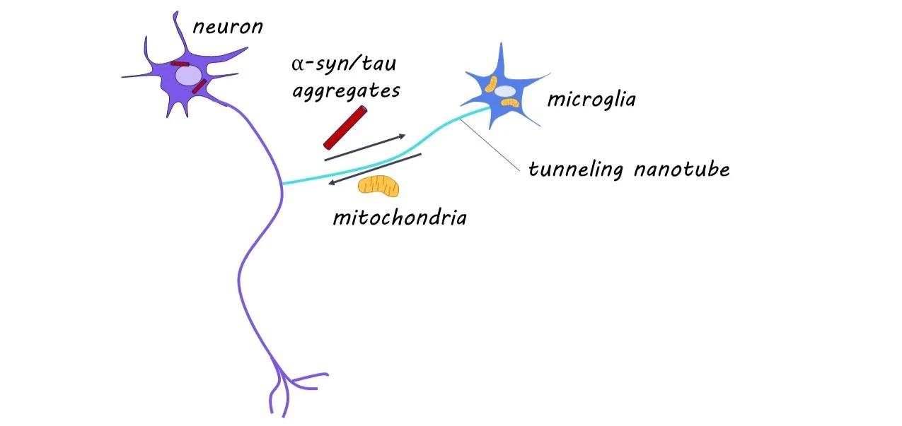
The buildup of protein aggregates in neurons is a feature of many neurodegenerative diseases, including Alzheimer’s and Parkinson’s disease. In this study, it was discovered that tunneling nanotubes connecting microglia and neurons is pivotal in the removal and degradation of these protein aggregates. 

What's the science?

While alpha-synuclein (a-syn) and tau are proteins that perform essential functions in healthy neurons, they can become disruptive and lead to neural damage when they accumulate in the brain. Microglia are immune cells present throughout the brain that clear protein aggregates present in extracellular space and help maintain healthy neural activity. Interestingly, microglia form open channels with neighboring cells, known as tunneling nanotubes (TNTs), to transport protein aggregates and healthy mitochondria (often referred to as ‘the powerhouse of the cell’) to maintain healthy neural function. Before this study, it was unknown whether microglia form TNTs to pathologic neurons to remove protein aggregates. 

This week in Neuron, Scheiblich and colleagues find that microglia form TNTs with pathologic neurons, to both remove protein aggregates and deliver healthy mitochondria and that these actions are critical for neural survival. 

How did they do it?

They performed RNA sequencing on neurons exposed to protein aggregates to compare how a-syn and tau aggregation affect neuron-microglia interaction. To assess mitochondria levels, they used a fluorescent dye taken in by mitochondria cells. These exposed neurons were cultured along with microglia, and a 3D laser scanning microscopy (a technique that allows reconstruction of 3d structures in the brain) was used to measure TNT formation and quantify the movement of protein aggregates and mitochondria between neurons and microglia. To understand the mechanism of protein transfer through TNTs, they knocked out different components of the Rac-PAK pathway (involved in responding to extracellular signals). 

To explore whether TNT formation occurred in human tissue as well, they quantified TNT formation in postmortem human brain tissue from patients with neurodegenerative diseases. The authors used Calcium imaging and patch-clamp experiments when exposed neurons were cultured with or without microglia to measure impairments in neural function. They fluorescently labeled mitochondria, to see whether mitochondria moved from microglia to neurons. They also used an inhibitor of mitochondria to determine whether mitochondria transfer from microglia to neurons was responsible for the improved function in neurons.

They also introduced genetic variants correlated with neurodegenerative diseases to see how these genetic alterations disrupted microglia and neuronal processes. 

What did they find?

Through RNA sequencing, they found 951 genes differentially regulated in protein aggregate-exposed neurons. These neurons exhibited reduced levels of mitochondria and increased markers of cell death. They found microglia to be pivotal in the removal of protein aggregates from neurons via TNTs, and that neurons alone cannot degrade protein aggregates. The TNTs formed between microglia and neurons contained protein aggregates, and this transport was found to be unidirectional from affected neurons to microglia. They demonstrate that the Rac-PAK pathway is critical in the removal of protein aggregates via TNTs. When analyzing postmortem human brain tissue, they found TNT formation between neurons and microglia in diseased samples. 

Using calcium imaging and patch-clamp methods, they found decreased neural activity in isolated exposed neurons and restored neural activity in exposed neurons cultured with microglia. When co-cultured, they found increased transfer of mitochondria to neurons, and this unidirectional transfer was correlated with improved function in affected neurons. This suggests that the neuroprotective effect microglia have on neurons with high protein aggregation is partly through the transfer of mitochondria via TNTs

They found that genetic disruption of genes associated with neurodegenerative diseases reduced the protein aggregate removal via TNTs and disrupted the transport of mitochondria from microglia to affected neurons. 

What's the impact?

This study illuminates the critical role of TNT connections between microglia and neurons, in both the clearance of protein aggregates from neurons and the transfer of mitochondria to effective neurons. The current findings open the door for much research into how this process may become disrupted in many neurodegenerative diseases such as Alzheimer’s and Parkinson’s disease. 

Access the original scientific publication here.

Why Are Vitamins Important for Your Brain?

Post by Meagan Marks

Nutrition and brain health

What you eat plays a crucial role in brain physiology and function and influences mood and mental health. Proper intake of micronutrients is vital for neurological and psychiatric well-being, with inadequate micronutrient levels potentially leading to problems with stress, hormone regulation, sleep, and overall cognitive function. 

What are vitamins?

Vitamins are essential organic compounds that our bodies need to function properly. The human body cannot make most vitamins on its own, so we must get them from our diet. When it comes to the brain, vitamins help maintain optimal psychiatric and neurological health by performing key functions: neurotransmitter synthesis, neuron growth, and neurochemical balance regulation. Particularly important, vitamins help protect against excitotoxicity, oxidative stress, and neuroinflammation – three main drivers of psychiatric and neurological disease.

Despite their importance, vitamin deficiencies are surprisingly common, particularly in the United States where the prevalent Western diet is highly processed and often lacking in micronutrients. In fact, it’s been suggested that this rise in micronutrient deficiency may be one factor related to the rise of mental and neurological disorders seen in the US.

So, which vitamins are indispensable for neurological functioning? How do they function in the brain? What risks come with deficiency, and what are some easily accessible food sources to add to your diet?

Vitamin A

Vitamin A is most known for its role in neurodevelopment and synaptic plasticity. It interacts with receptors in the brain that regulate the expression of genes that are involved in the formation of new neurons and neural connections. This is essential for learning, memory, and recovery from brain injuries. In addition, vitamin A also holds antioxidant properties that help protect the brain against oxidative stress and damage.

Adequate vitamin A levels are associated with better cognitive function, while deficiencies have been linked to cognitive impairments and increased risk of neurodegenerative disease. Food sources high in vitamin A include mangos, papayas, leafy vegetables, milk, eggs, and cheese. A note of caution, however: too much vitamin A (typically via oversupplementation) can be toxic!

B Vitamins

The B vitamins are a group of eight vitamins that perform some of the most essential and interconnected roles in the brain. B vitamins predominantly act as coenzymes, fusing with numerous proteins to boost their potential and help them perform more chemical reactions. Most notably, the B vitamins are responsible for synthesizing neurotransmitters and other signaling molecules in the brain.

Specific examples of their functions include:

B1 (thiamine) synthesizes precursors to neurotransmitters and other compounds essential to neuron function and structure. This includes acetylcholine, a neurotransmitter important to memory, learning, arousal, and attention.

B2 (riboflavin) acts as an antioxidant and helps regulate thyroid hormones that are essential to metabolism. B2 also helps to form B6, an extremely important B vitamin listed below.

B3 (niacin) also holds antioxidant properties, as well as helps with DNA metabolism and repair, cellular signaling, and oxidative reactions.

B5 (pantothenic acid) aids in neuron communication by myelinating axons and contributing to the structure of cells.

B6 is one of the most important vitamins to brain health. It is required to make the “feel good” neurotransmitters dopamine and serotonin, as well as the inhibitory neurotransmitter GABA. With even a mild B6 deficiency, levels of these neurotransmitters can drop in the brain. This leads to an excess of the excitatory neurotransmitter glutamate, which is linked to anxiety, depression, and excitotoxicity.

B8 (folic acid) and B12 are also both important to neurotransmitter synthesis, while B7 (biotin) is important for glucose metabolism.

Deficiencies in any of these vitamins will alter the brain’s ability to produce energy and perform essential processes, which can lead to chronic disease, psychiatric illness, and cognitive decline. Insufficient intake can manifest in many forms, including irritability, disordered sleep, inflammation, dementia, depression, and mental fog. Good sources of the B vitamins include fish, nuts, brown rice, pasta, citrus fruits, and leafy greens. 

Vitamin C

Vitamin C enhances neuron communication by myelinating axons, wrapping them in a protective, lipid-rich blanket. It also acts as a cofactor in producing dopamine and norepinephrine, neurotransmitters essential for motivation, mood, sleep, attention, and stress. Vitamin C is also a strong antioxidant, protects against excitotoxicity, and reduces inflammation by removing the proteins that cause it.

Deficiencies in vitamin C can lead to neuroinflammation, excitotoxicity, and oxidative stress, and may manifest as fatigue, mood swings, irritability, and depression. Great sources include bell peppers, citrus fruits, and various greens and vegetables.

Vitamin D

Vitamin D is well-known for managing fetal neurodevelopment and dopamine regulation. During pregnancy, vitamin D deficiency has been linked to abnormal neurodevelopment and is associated with an increased risk of schizophrenia and autism, which is thought to be due to impacts on dopamine regulation. Vitamin D supports the synthesis, transportation, and release of dopamine and is also positively correlated with growth and population of dopaminergic neurons.

Having a deficiency in vitamin D can increase the risk of cognitive decline, Alzheimer's disease, and Parkinson's disease and can greatly imbalance dopaminergic signaling. Great sources include fish, butter, cod liver oil, meat, milk, and eggs.

Vitamin E

Vitamin E is another vitamin that plays a strong protective role against oxidative stress, excitotoxicity, and neuroinflammation. Vitamin E can directly break down glutamate in the brain, reducing the risk of excitotoxicity and preventing cell damage. It also lowers levels of inflammatory proteins, reducing brain inflammation. Vitamin E also protects brain tissue by serving as an antioxidant.

Deficiencies may result in cognitive impairments, motor issues and muscle weakness. Good sources of vitamin E include seeds, nuts, avocados, sweet potatoes, and fish.

Vitamin K

While vitamin K is mostly known for its role in blood clotting and bone health, some emerging evidence suggests it also plays a role in brain function. Current findings suggest that it can reduce oxidative stress and neuroinflammation, help maintain cell membrane integrity, and support neuron survival and growth, but more research is needed to fully understand its role. Past studies have also shown that sufficient levels of vitamin K may improve cognitive performance and slow cognitive decline. 

Deficiencies may result in fatigue, irritability, and sleep disturbances, and great sources include leafy greens, vegetable oils, fruits, meat, eggs, and soybeans. 

What’s the takeaway?

Proper nutrition is essential to optimal neurological and psychiatric functioning. Consuming proper levels of micronutrients, whether through diet alone or with the help of supplementation under medical supervision, has been shown to 1) reduce stress, anxiety, and depression, 2) protect against inflammation, oxidative stress, and excitotoxicity, and 3) improve memory and cognitive tasks. While nutritional neuroscience is still a growing field, there is evidence that a proper and personalized diet could help prevent, manage, or even treat neurological and psychiatric disorders.

Access topic overview citations here: 1, 2, 3, 4, 5, 6, and 7.