Identifying Targets for Neuromodulation of PTSD

Post by Kelly Kadlec

The takeaway

In this study, the authors investigated the functional connectivity between brain regions of veterans with and without brain lesions to identify a neural circuit for Post-traumatic stress disorder (PTSD). They demonstrated the potential therapeutic benefits of targeting brain regions within this circuit using noninvasive neuromodulation.  

What's the science?

Post-traumatic stress disorder can result from the experience of a traumatic event or series of events and symptoms can include anxiety and depression. Despite its prevalence, traditional medication and psychotherapy treatments are often not effective, leading researchers to explore neuromodulation techniques such as transcranial magnetic stimulation (TMS). Unfortunately, one of the most commonly implicated regions in PTSD, the amygdala, is not accessible by TMS. Therefore, there is a need to identify alternative modulation targets. Recently in Nature Neuroscience, Siddiqi and colleagues investigated circuits involved in  PTSD in veterans and identified and demonstrated the medial prefrontal cortex (mPFC) as a potential target for TMS treatment of PTSD.

How did they do it?

First, the authors compared rates of PTSD in veterans with and without penetrating traumatic brain injury (TBI), and examined which lesioned brain areas were most associated with reduced rates of PTSD. Additionally, they used functional MRI (fMRI) to determine the functional connectivity (FC) of brain areas where lesions seemed to protect against PTSD. This connectivity was established by lesion network mapping, which relies on the connectome database of resting-state fMRI for 1,000 individuals. Then, they compared the patterns of FC found in veterans with TBI to those without TBI including a large cohort of veterans with PTSD.   

The authors then assessed whether neuromodulation of implicated brain regions resulted in changes in PTSD symptoms. First, they evaluated how TMS in regions identified by the connectivity results compared with TMS in other regions in terms of the ability to relieve PTSD symptoms. In addition, they evaluated how different types of modulation (i.e. inhibitory, excitatory) impacted PSTD symptoms.

What did they find?

First, the authors report that veterans with penetrative brain injuries had reduced rates of PTSD. In particular, individuals with damage to the amygdala were the most protected against developing PTSD. 

Next, the authors found that reduced functional connectivity between the mPFC, amygdala, and hippocampus, was associated with reduced PTSD symptoms. This ‘lesion-derived PTSD circuit’ was also validated in veterans without TBI by comparing FC in individuals with and without PTSD. They were able to further examine this circuit in individuals who had received TMS for PTSD in a previous study and found that, as hypothesized, a reduction in these patient’s symptoms was associated with a reduction in FC between the areas in this circuit. 

Finally, the authors validated the neural circuit they identified as a target for TMS-based treatment of PTSD by showing that TMS in regions within the circuit was more effective than TMS in other areas in reducing symptom severity. In addition, as predicted by the FC results, applying inhibitory modulation to these regions resulted in a decrease in PTSD symptom severity while applying excitatory modulation had the opposite effect.  

What's the impact?

PTSD can have a negative impact on the quality of life of individuals afflicted and current treatments are unfortunately limited in their efficacy. The findings in this study demonstrate the mPFC as a promising target for non-invasive neuromodulation and reduction of PTSD symptoms. Further, the results of this study present causal evidence for a critical neural circuit in PTSD.  

Access the original scientific publication here.

Can Alzheimer’s disease be prevented?

Post by Shireen Parimoo

Overview

Alzheimer’s disease (AD) is a neurodegenerative disorder that affects millions of older adults globally, with five million new cases every year. In the brain, AD is characterized by the accumulation of amyloid beta plaques and neurofibrillary tangles made up of misfolded tau proteins. These plaques and tangles accumulate in brain areas like the hippocampus that are important for memory, eventually leading to cell death and atrophying of affected regions. As a result, common cognitive impairments in AD include memory problems, diminished attentional and decision-making capacity, and loss of language abilities. Alzheimer’s disease also impacts mental health and day-to-day functioning as patients experience problems with sleep, depression, apathy, aggression, and psychosis. Although there is no known cure for AD, treatments currently exist to alleviate some of the cognitive symptoms of the disease.

Risk Factors for Alzheimer’s disease

One of the major risk factors for AD is genetic susceptibility. Normally, we inherit one copy of a gene - called an allele - from each parent.  A small subset of individuals with AD have early-onset or familial AD that they develop due to inherited gene mutations from one or both of their parents. Symptoms of early-onset AD generally begin in the 30s and 40s and quickly lead to deteriorating health and well-being. For most patients, however, symptoms of AD emerge later in life, around 65 years old. In these cases of sporadic AD, the apolipoprotein E (APOE) has been identified as a key genetic risk factor. Specifically, having one or two copies of the APOE-4 allele increases the risk of AD because it leads to higher levels of amyloid plaques in the brain. 
 
Besides genetic risk, many lifestyle and medical factors can also increase the risk of developing AD. For example, hypertension and elevated cholesterol levels are both associated with a higher risk of developing AD, as are obesity and diabetes. Research studies have also identified smoking, poor sleep, social isolation and loneliness, and chronic stress as lifestyle predictors of AD and cognitive decline later in life. Fortunately, many of these risk factors are modifiable, which means that it is possible to make lifestyle changes to lower the odds of developing AD.

The Importance of Lifestyle // Protective Factors

Protective factors are variables that - as the name suggests - protect against the risk of developing AD. As with risk factors, protective factors can be immutable or modifiable. A genetic protective factor, for instance, is the presence of two copies of the APOE-2 allele. Unlike APOE-4, the APOE-2 variant of the gene reduces the risk of developing AD. Interestingly, although genetic risk itself is not currently modifiable, studies show that there is an interaction between genetic and lifestyle factors, or in other words, between nature and nurture.
 
Fortunately, research studies over the past few decades have identified several modifiable lifestyle factors that have a protective effect against dementia and cognitive decline, even if there is a genetic risk for developing AD. This means that we can take measures to improve our brain health into our own hands and, at the very least, mitigate the rate of cognitive decline in older age.

  1. Physical activity is an all-around protective factor against multiple conditions including AD and dementias. Exercising reduces blood pressure and proinflammatory activity, both of which are risk factors for AD. In the long term, physical activity also improves blood flow to the brain, which is important for brain health and functioning in general. Aerobic exercises like running, walking, and cycling increase neurotrophic factors in the brain, which are proteins that promote the growth, plasticity, and survival of neurons. The effects of exercise on the hippocampus in particular are well-documented. Both healthy older adults and AD patients who engage in physical activity have higher levels of neurotrophic factors and in some cases, larger hippocampal volume because of exercise. Older adults at risk for AD who engage in physical activity also show less hippocampal atrophy, which could slow down the onset of dementia and memory-related symptoms.


  1. Diet and nutrition are also important for maintaining brain health and protecting against cognitive decline. Sources of unsaturated fats like olive oil and nuts are important for helping neurons maintain the integrity of their synapses (i.e., gaps between neurons). Antioxidants like folic acid from fruits and vegetables, as well as vitamins like vitamin D confer some protection against neurodegeneration by regulating neurotrophic factors to maintain neuronal health and by clearing amyloid protein in the brain. In fact, individuals with a vitamin D receptor gene mutation tend to be more at risk for developing AD. Thus, a well-balanced and nutritious diet - such as the Mediterranean diet, which is rich in antioxidants - can reduce the risk of developing AD in older age.


  1. Psychosocial factors also have a protective effect against dementia, including social enrichment, educational attainment, leisure activities, and psychological well-being. Older adults with an active social life are more likely to be intellectually and socially stimulated, which is linked to increased neurogenesis and lower levels of stress. Similarly, having a strong social support system increases our sense of belonging, reduces feelings of loneliness, and improves our mental well-being. Higher educational attainment early in life and stimulating leisure activities - like learning a new language or playing an instrument - also help prevent the onset of dementia. The idea is that engaging in these social and intellectual domains ensures that the neural circuits in the brain remain active and are less likely (or at the very least slower) to deteriorate during aging.


  1. Sleep disturbances are linked to a higher risk of cognitive decline and AD. For example, amyloid and tau accumulation and clearance are modified by sleep patterns. Getting enough good quality sleep is important because that is when toxins like amyloid and tau are cleared through the glymphatic (fluid flow) system in the brain. Interestingly, the relationship between sleep and AD appears to be bidirectional: healthy individuals with amyloid and tau pathology (precursors to AD) and individuals with AD tend to have poor sleep quality and more sleep disturbances. As a result, it is unclear whether sleep quality contributes to the onset of AD, whether it is one of the outcomes of AD pathology, or some mix of the two.

 
Overall, there are many modifiable lifestyle variables that can help prevent cognitive decline and protect against the onset of neurodegenerative disorders like AD. Emerging research indicates that a personalized, multidomain, lifestyle-based interventional approach will have the most beneficial effects on slowing down pathological processes in aging, especially in older adults who might be genetically at risk for developing dementia.

References +

Li et al. (2014, BioMed Research International). Behavioral and psychological symptoms in Alzheimer’s disease. Link: https://www.ncbi.nlm.nih.gov/pmc/articles/PMC4123596/

Silva et al. (2019, Journal of Biomedical Science). Alzheimer’s disease: Risk factors and potentially protective measures. Link: https://jbiomedsci.biomedcentral.com/articles/10.1186/s12929-019-0524-y

Wang et al. (2021, Alzheimer’s & Dementia). Shared risk and protective factors between Alzheimer’s disease and ischemic stroke: A population-based longitudinal study. Link: https://alz-journals.onlinelibrary.wiley.com/doi/full/10.1002/alz.12203

Iso-Markku et al. (2021, British Journal of Sports Medicine). Physical activity as a protective factor for dementia and Alzheimer’s disease: Systematic review, meta-analysis, and quality assessment of cohort and case-control studies. Link: https://bjsm.bmj.com/content/bjsports/56/12/701.full.pdf

Rosenberg et al. (2020, The Journal of Prevention of Alzheimer’s Disease). Multidomain interventions to prevent cognitive impairment, Alzheimer’s disease, and dementia: From FINGER to World-Wide FINGERS. Link: https://link.springer.com/content/pdf/10.14283/jpad.2019.41.pdf

Walsh & Brayne (2021, Alzheimer’s & Dementia). Does playing a musical instrument prevent dementia? Link: https://alz-journals.onlinelibrary.wiley.com/doi/abs/10.1002/alz.049684

Liu et al. (2013, Nature Reviews Neurology). Apolipoprotein E and Alzheimer’s disease: Risk, mechanisms, and therapy. Link: 10.1038/nrneurol.2012.263

Qiu et al. (2009, Dialogues in Clinical Neuroscience). Epidemiology of Alzheimer’s disease: Occurrence, determinants, and strategies toward intervention. Link: https://www.ncbi.nlm.nih.gov/pmc/articles/PMC3181909/

Kuiper et al. (2015, Ageing Research Reviews). Social relationships and risk of dementia: A systematic review and meta-analysis of longitudinal cohort studies. Link: https://individuallytics.com/wp-content/uploads/2020/02/Social-Relationships-and-Risk-of-Dementia-Kuiper-et-al-2015.pdf

Wang et al. (2017, PLoS Medicine). Association of lifelong exposure to cognitive reserve-enhancing factors with dementia risk: A community-based cohort study. Link: https://journals.plos.org/plosmedicine/article/file?id=10.1371/journal.pmed.1002251

Lucey, B. P. (2020, Neurobiology of Disease). It’s complicated: The relationship between sleep and Alzheimer’s disease in humans. Link: https://www.sciencedirect.com/science/article/pii/S0969996120303065

Wang & Holtzman (2020, Neuropsychopharmacology). Bidirectional relationship between sleep and Alzheimer’s disease: Role of amyloid, tau, and other factors. Link: https://www.nature.com/articles/s41386-019-0478-5

Zhang et al. (2022, Translational Psychiatry). Sleep in Alzheimer’s disease: A systematic review and meta-analysis of polysomnographic findings. Link: https://www.nature.com/articles/s41398-022-01897-y

Irwin & Vitiello (2019, The Lancet Neurology). Implications of sleep disturbance and inflammation for Alzheimer’s disease dementia. Link: https://www.thelancet.com/journals/laneur/article/PIIS1474-4422(18)30450-2

How Neurons in the Human Brain Encode the “When” of Experiences

Post by Meagan Marks

The takeaway

Neurons in the medial temporal lobe help distinguish temporal patterns within recurring experiences, influencing the implicit prediction of future events and guiding subsequent behavior.

What's the science?

Whether performing a daily routine or learning a new task, the human brain is constantly integrating aspects of experience—specifically, the “what” (objects/events), “where” (spatial location), and “when” (temporal structure)—to create a cohesive understanding of the world. 

Temporal structure—the “when” of an experience—allows the brain to organize information based on order in time. Encoding information in such a manner is crucial to memory formation, sequential learning, and decision making. By recognizing subtle sequences and patterns in time, the brain can predict future outcomes and adapt behavior accordingly. This process is needed for everyday tasks, like comprehending the words on a book page or responding to the flow of ideas during a conversation.  

Temporal structure is vital for survival and success, but the specific neuronal mechanisms behind this cognitive process remain unclear. This week in Nature, Tacikowski and colleagues explore how neurons encode the temporal structure of experiences into memory by observing the neural activity of human participants. 

How did they do it?

To determine how human neurons encode temporal structure, the authors recruited 17 participants, all of whom had intracranial electrodes implanted for clinical reasons. The authors then recorded the activity of individual neurons within the medial temporal lobe (MTL)—a region of the brain known for its role in memory and spatial cognition. A complex behavioral task was also created to ensure that subtle temporal patterns were presented to the participants as neuronal activity was recorded.   

The behavioral task consisted of three phases. During the first, participants were repeatedly shown six images of people that elicited a strong response from neurons in the MTL. These images were displayed at random and participants were asked to identify the gender of each person shown. 

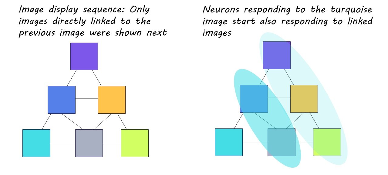
During the second phase, the same six images were repeatedly presented over the course of six trial rounds, but this time in a predetermined sequence. This sequence was based on a pyramidal structure established behind the scenes, where one image was assigned the top of the pyramid, two were assigned to the middle, and three were assigned to the bottom. Although the participants were only shown one image at a time, these images were shown in alignment to this pyramidal arrangement, so that only images “touching” each other on the pyramid could follow each other in the display line up. During these trials, the participants were engaged in a separate task—determining if the images had been flipped—and were not aware of the display pattern. 

The third and final phase mimicked the first, however, the participants had been sufficiently exposed to the display sequence. 

What did they find?

During the first phase of the behavioral experiment, the researchers noticed that certain neurons in the MTL exhibited a significantly stronger response to one particular image compared to the others. As the experiment progressed, these neurons adjusted their activity based on the display pattern. With continued exposure, some of the initially selective neurons began responding more strongly to images that were connected on the pyramid to the original image they had favored (i.e., images connected within the sequence). This strengthening of neuronal activity directly aligned with exposure to the pattern, indicating that these adaptable neurons made associations between images and embedded the sequence into memory. The sequence was even ‘replayed’ by the neurons spontaneously during study breaks, strengthening the memory of the pattern. These neurons were distributed across the hippocampus and entorhinal cortex, suggesting that the hippocampal-entorhinal system plays a key role in encoding temporal structure.

Participants also exhibited delayed behavioral responses in the final phase when an image broke the learned sequential pattern. Given that participants remained unaware of the display pattern, it can be inferred that their recognition and memorization of the sequence occurred largely implicitly.

What's the impact?

This study is the first to show that neurons in the hippocampus and entorhinal cortex work together to encode the temporal structure of experience. These neurons play a crucial role in recognizing and memorizing patterns, influencing the implicit prediction of future events, and guiding subsequent behaviors. These findings offer a deeper understanding of the neuronal mechanisms that underlie human behavior and experience.