The Role of Mitochondria in Age-Related Cognitive Decline

Post by Kelly Kadlec

The takeaway

This study investigated how mitochondria influence cognitive decline related to aging. In addition to illuminating the molecular link between synaptic excitation and mitochondrial gene transcription, the authors demonstrate how this molecular cascade could provide a basis for treatments to improve age-related cognitive decline.      

What's the science?

A loss of energy as we age is a nearly universal experience, and a decline in cognitive function is seen as largely inevitable. It is thought that this change in the aging brain is related to changes in mitochondrial function, but the molecular underpinnings of this process have remained largely unknown. Understanding the relationship between neuronal activity and mitochondrial DNA transcription may provide key insights into the aging brain and how we might counteract functional decline by developing treatments that target this interaction. Last week in Science, Li and colleagues uncovered the molecular cascade linking synaptic excitation and mitochondrial DNA transcription and demonstrated that targeting this cascade can improve age-related cognitive decline in rodents.  

How did they do it?

The authors investigated the molecular process of activity-dependent mitochondrial DNA transcription in mice using a broad range of in vivo and ex vivo techniques. First, the authors used RNAscope and optogenetics in hippocampal brain slices along with foot-shocks and quantitative real-time PCR in vivo to establish that neuronal activity interacts with mitochondrial transcription. Then, they compared this mitochondrial expression in young and aged mice. To better understand the cause of the age-related changes they observed, they again used optogenetic and pharmacological tools to isolate a critical role for activity-dependent calcium. 

Next, they conducted immunogold electron microscopy in the hippocampus to determine whether or not this calcium dependency is regulated by calmodulin-dependent protein kinase II (CAMKII). The authors then sought to determine whether mitochondria can decode calcium activity through CRE-like sequences. They used a DNA-affinity assay to identify the presence of mitochondrial CREB and derived CREB activity sensors to directly probe its function. 

Finally, the authors used whole-cell recordings, intracellular ATP measurements, and a variety of genetic techniques to measure and modulate neuronal activity. They examined the role that activity-coupled mitochondrial transcription plays in synaptic function and regulation. They tested these findings under the hypothesis of age-related changes by investigating how inhibiting or enhancing activity-dependent mitochondrial transcription impacts association-based learning in mice of different ages.

What did they find?

The authors first show a causal coupling between neuronal and synaptic excitation and mitochondrial DNA transcription. This expression was reduced in aged mice compared to young mice and was also associated with lower levels of activity-dependent mitochondrial calcium. The authors subsequently found that activity-coupled mitochondrial transcription relies on mitochondrial calcium.

The authors also probed the mechanisms that link neural activity with mitochondrial transcription and found that this process recruits the same molecules that have an established role in activity-transcription coupling in the nucleus. Specifically, activity-dependent mitochondrial transcription and calcium were regulated by CaMKII. Moreover, the translation from activity-dependent calcium to DNA transcription is mediated by mitochondrial CREB.

The authors also show how activity-coupled mitochondrial transcription regulates both synaptic and mitochondrial resilience, further demonstrating how this molecular process mediates both neuronal energy reserves and memory processes. Finally, they show that restoring activity-dependent mitochondrial transcription in aged mice enhances memory, suggesting a mitigation of age-related cognitive decline.

What's the impact?

Activity-dependent mitochondrial DNA transcription has long been suspected to play a critical role in maintaining neural energy reserves, and this study provides key insight into the molecular cascade underlying this process. The authors also show age-related alterations in this pathway that likely contribute to cognitive decline during aging. The authors also demonstrate that reinvigorating this pathway may reduce or even reverse age-related decline in brain function.  

Access the original scientific publication here.

How Experiencing a Negative Event Alters Responses to Others

Post by Shahin Khodaei

The takeaway

Previous negative experiences affect how mice respond to other mice in a similar state. This behavior is mediated by the corticotropin-releasing factor (CRF) system in a region of the brain called the medial prefrontal cortex.

What's the science?

As humans, we recognize the signs that another person has experienced a stressful event and respond to them, for example showing consoling or prosocial behaviors. Similar to humans, the previous experiences of a mouse can affect how it responds to another stressed mouse. These different responses may be regulated by the corticotropin-releasing factor (CRF) system in the brain, specifically in a region called the medial prefrontal cortex which is important in emotion and socialization. This week in Nature Neuroscience, Martese and colleagues investigated 1) the ways in which mice with different experiences react to a stressed mouse, and 2) the role of the CRF system in these responses. 

How did they do it?

The authors used mice as either observers or demonstrators. Observer mice were placed in a box and presented with two demonstrator mice – one that was given a stressful experience right before the test (either being restrained for 15 minutes, or getting a shock to their feet), and one that was unstressed. The authors looked at how the observer interacted with the demonstrators. In some experiments, the observer was subjected to a stressful “negative self-experience” (NSE) one day before the test. The authors compared how much the observer interacted with each of the demonstrators. 

To study whether CRF neurons in the medial prefrontal cortex played a role in the behavior of the observers, the authors used genetic techniques to add certain genes to these brain cells. These genes either 1) decreased the level of CRF, 2) caused the cells to emit light when they were activated, which could then be measured using a microscope, or 3) allowed the authors to decrease the activity of the cells using optogenetics.

What did they find?

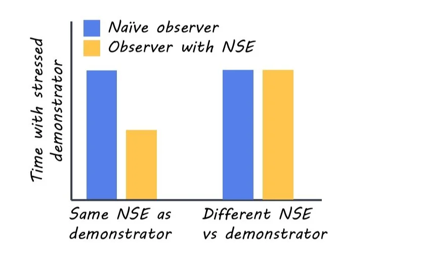
When a naïve observer was placed in front of one naïve and one restraint-stressed demonstrator, the observer spent more time sniffing and interacting with the stressed demonstrator. In contrast, if observer mice had experienced a NSE of restraint, they spent less time with the restraint-stressed demonstrator (and sometimes completely avoided them). This change in response was experience-specific. If the type of stressful experience did not match between the observer and demonstrator (i.e., one was footshock-stressed and the other was restraint-stressed), the observer with NSE behaved just like a naïve observer, and spent more time with the stressed demonstrator. This suggests mice were responding uniquely to other mice experiencing similar negative events. For male mice, the behavior of the observer with NSE depended on the social status of the mouse: dominant mice avoided the stressed demonstrator, and non-dominant mice showed no preference between the stressed and naïve demonstrator. For female mice, the behavior of the observer with NSE depended on the phase of the estrus cycle (the mouse equivalent of the menstrual cycle). 

The authors focused on the CRF system in the medial prefrontal cortex. They showed that reducing the level of CRF in these neurons made observers with NSE behave more similarly to naïve observers. The authors then assessed how the activity of these neurons changed in observers. They found that in naïve observers, the activity of CRF neurons increased when they were near the un-stressed demonstrator. In contrast, for observers with NSE, the activity of these neurons increased near the stressed demonstrator. If they reduced this CRF neural activity using optogenetics, naïve observers started to avoid stressed demonstrators (i.e., behave more like NSE observers) and NSE observers preferred stressed demonstrators (i.e., behave more like naïve observers). These results show that the CRF system is involved in how previous experiences influence the way mice approach stressed animals. 

What's the impact?

This study sheds light on the mechanisms involved in how previous experiences influence behavioral responses to stress in others. Importantly, this research reveals a neurobiological mechanism involving the CRF system for how interactions with others may differ, based on previous negative experiences.

Access the original scientific publication here.

“Starter Cells” Mediate Stress-Induced Depression-Like Behaviors

Post by Amanda Engstrom 

The takeaway

In this study, researchers identified a small population of stress-responsive neurons, termed “starter cells”, in subregions of the hypothalamus-habenula circuit. These neurons are required for the development of depression-like behaviors in mice. 

What's the science?

Chronic stress has long been considered a major risk factor for depression. Identifying specific neurons that respond to stress could offer insights into anti-depressant interventions. Experience-responsive neuronal ensembles are small populations of neurons that are functionally involved in distinct experiences. The lateral habenula (LHb) and the lateral hypothalamus (LH), which projects into the LHb, have been implicated in encoding aversion and depression-like behaviors during chronic stress. However, the core stress-responsive neurons within this circuit have not been identified. This week in Neuron, Zheng and colleagues identified a small population of neurons within the LH-LHb circuit, investigating their role in mediating depression-like behaviors due to stress. 

How did they do it?

The authors combined c-fos immunostaining (a marker for neuronal activity) and a Robust Activity Marking (RAM) system to identify the functional neurons within the LH-LHb circuit that respond to stress. RAM is a viral strategy to capture and label experience-responsive neurons in mice. Using this approach, the mice were exposed to various types of stress, such as restraint stress, and the authors determined which cells were activated in response to stress. They then used a chemogenetic strategy where they expressed a compound to inhibit the identified neurons while the mice were exposed to stress. This enabled them to determine the effect on depression-like behaviors when these neurons were nonfunctional. In a complementary experiment, the authors used photostimulation, which uses light to activate these same cells to mimic the activity of the neurons in the absence of a stressor and determine if the mice would display depressive-like behaviors. To characterize the synaptic connections from the LH stress-responsive neurons to LHb subpopulations the authors used optogenetics - assisted electrophysiology, a technique that uses optical and genetic manipulation to control neuronal activity while simultaneously recording electrical activity. Finally, they visualized the process of neuronal recruitment by labeling stress-responsive LHb neurons via RAM at different stages of chronic stress. 

What did they find?

After exposing mice to stress the authors identified a small population of neurons in the LHb and the LH (10% and 5% respectively) that were activated and labeled via the RAM system as well as c-fos positive. In both the LH and the LHb the active cells localized to subregions, specifically the middle part of the lateral hypothalamus (mLH) and the medial part of the lateral habenula (LHbM). Chemogenetic inhibition of the stress-responsive mLH or the LHbM neurons during chronic stress stopped the development of depression-like behaviors in mice. Additionally, activating these neurons via photostimulation is sufficient to cause depression-like behaviors. These data suggest that both regions are required for the development of depression-like behaviors caused by chronic stress. Using optogenetics-assisted electrophysiology, the authors showed that the stress-responsive neurons in the mLH and LHbM formed dominant excitatory connections after one exposure to stress, and they are selectively potentiated during chronic stress. The authors termed these cells “starter cells”. LHbM starter cells propagate hyperactivity across the entire LHb via local excitatory connections. Finally, the authors visualized the progressive recruitment of LHb neurons over time due to chronic stress. Initially, very little fluorescence signified very few stress-induced neurons, but they gradually increased over time. This indicates that initially stress-irresponsive LHb neurons were gradually becoming responsive, and the intensity of this response increased over time. The LHb stress-responsive neurons were initially limited to the LHbM and gradually spread throughout the entire LHb. 

What's the impact?

This study identified a small population of stress-responsive neurons in subregions of the lateral hypothalamus (LH) and LH lateral habenula (LHb) that are critical for stress-induced depression-like behavior in mice. The authors identified and characterized a novel core functional unit within the larger LH-LHb circuit, further highlighting the importance of ensemble sparsity (a small number of functional cells within a larger ensemble) in various brain functions. Moreover, by identifying stress-responsive neurons, these data offer new insights into potential targets for antidepressant therapies.

Access the original scientific publication here.