Can Combining Brain Imaging Techniques Help Us “See” Our Thoughts?
Post by Christopher Chen
The takeaway
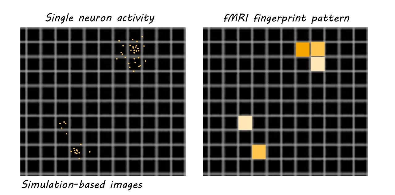
Increasing evidence from brain imaging studies suggests that fingerprint-like patterns of brain activation may reflect information during cognitive processes like emotion and perception. Researchers think that these patterns may represent sparse code at the neuronal level - reproducible patterns of neural firing in a small percentage of neurons.
What underlies perception, cognition and emotion?
Even with the advent of modern brain imaging techniques in the late twentieth century, researchers have still not fully answered a question dating back to ancient Greece and Aristotle’s musings on the mind: what is the basis of human thought?
That said, science has made great progress in painting the big picture regarding the brain’s role in discrete tasks. For example, viewing an image of a face elicits distinct activity patterns in a region of the brain called the fusiform gyrus, and thinking about the connection between two events is related to activity in a brain region called the hippocampus. Showing that specific tasks stimulate specific brain regions, however, is not quite the same as determining how the brain processes information.
Recently in Neuroimage, Jaaskelainen and colleagues offer evidence that combining two techniques – functional magnetic resonance imaging (fMRI) and two-photon calcium imaging (2PCI) – will help researchers demonstrate that the brain processes information through the activation of specific populations of neurons. Combining these two techniques could potentially help neuroscientists take a huge step forward toward uncovering a biological foundation for human thought.
Fingerprint patterns of fMRI brain activity
Since its invention over thirty years ago, fMRI – a medical imaging technique measuring changes in the brain’s blood flow – has been critical in showing how specific stimuli like a human face, for example, activate specific brain regions. Since the early 2000s, a technique called multi-voxel pattern analysis (MVPA) has helped enhance fMRI studies by providing greater resolution in differentiating activity across brain regions, allowing researchers to describe stimulus-specific “fingerprints” in our brains. From these studies, we have determined that these fingerprints most likely represent information gleaned during tasks and presentation of various stimuli, an enormous victory in the quest to decode how our brains process information. Studies pairing fMRI and MVPA have generated a lengthy trail of evidence suggesting fingerprint patterns are linked to information representation in nearly all brain regions and helped establish fingerprints as a reliable biological representation of information in the human brain.
What is sparse neural code and why does it matter?
Studies in animals have been able to probe even deeper than fMRI studies. While fMRI measures the activity of brain regions, 2PCI can measure the activity of neurons within these brain regions by measuring neuronal calcium levels. These findings also highlight how efficient the brain can be when representing information, with one of the most notable 2PCI experiments showing that activation of only a minuscule (~0.5%) population of visual cortex neurons is required for visual processing in a monkey. This limited activation of specific neuronal populations in response to stimuli is aptly called sparse distributed representation, or sparse code.
Some believe sparse code is another level of detail in the journey to define the biological basis of cognition. Thus, some neuroscientists believe translating 2PCI sparse coding to fMRI fingerprints will help uncover how neuronal populations underlie the basis of thought formation.
What’s the relationship between fingerprints and sparse neural code?
Determining whether 2PCI-generated sparse coding underlies fMRI fingerprints is not an easy task. One of the primary questions is whether fMRI has the spatial resolution to record sparse representation. In other words, can fMRI which usually measures regional activity in millimeters, capture activity happening at the micrometer (1000-fold smaller) level? Evidence does suggest that the primary readout of 2PCI – calcium signaling – scales with the primary readout of fMRI, hemodynamic signaling, suggesting that fMRI can at the very least indirectly capture micrometer-level activity. Additionally, there is evidence from fMRI data suggesting a sparse coding-like distribution activity pattern in visual areas, which corresponds to findings from the visual area in 2PCI studies. And perhaps the most compelling evidence suggesting a functional similarity between sparse coding and fingerprints comes from a simulation experiment the authors performed, that generated amplified 2PCI data resembling an fMRI fingerprint. This evidence is a promising first step in a fuller characterization of just how intimately linked sparse coding and fingerprints are.
What does the future hold?
While still speculative, the idea that 2PCI sparse coding may inform fMRI fingerprints is enormous in scope. If true, it would show that the activation of very distinct, small populations of neurons lie at the core of basic human functions like cognition and emotional processing. A critical step, of course, is to measure the integration of sparse codes from multiple brain regions in higher-order cognitive tasks such as decision-making and learning. Experimentally, using both 2PCI and fMRI imaging in parallel will be instrumental in determining the similarities between sparse coding and fingerprint patterns. Studies could also employ a sparse coding amplification technique like the one just described to predict how well 2PCI data from animals scales with fMRI data in humans. Whether these studies reveal the neural basis of human cognition remains to be seen, but they will undoubtedly teach us more about how the interplay between sparse coding and fingerprints informs human behavior.

