Self-Modulation of Motor Cortex Activity after Stroke
Post by Elisa Guma
The takeaway
Self-modulation of the motor cortex using neurofeedback training in stroke patients enhances the activity of the affected motor cortex within a training session, but not long term. It also decreases brain white matter asymmetry in the corticospinal tracts detected 1 week after training.
What's the science?
Stroke survivors often face significant motor impairments, which can negatively affect their quality of life. Recent efforts have focused on rehabilitating the brain to improve motor function, rather than the muscles directly, with a particular focus on trying to decrease lateralization of motor cortex activity (i.e., relying less heavily on the unaffected hemisphere). This week in Brain, Sanders and colleagues investigate a novel therapeutic approach in which participants learn to self-modulate brain activity through real-time functional magnetic resonance imaging (fMRI) neurofeedback, which has shown promise in other disorders with aberrant brain activity patterns.
How did they do it?
The authors conducted a double-blind, randomized controlled trial on 24 chronic stroke survivors experiencing motor impairments in their upper limbs (mild to moderate). An initial baseline session was conducted to assess upper limb motor function and to acquire structural and functional magnetic resonance images of the brain. Following a baseline session, participants were equally and randomly split into real (n=12) or sham (n=12) neurofeedback training sessions lasting around 20 minutes for 3 days, with a 24 and then 48-hour break between each session. Participants returned for a 1-week and 1-month follow-up for behavioural assessments and brain imaging.
During the neurofeedback training sessions, participants were asked to lie in the fMRI scanner to measure brain activity while they opened and closed their hands (either left or right). Participants were shown two bars - red for the stroke-affected hand, and blue for the non-affected hand - on a screen representing neural activity within the hand knob region of the sensorimotor cortex. The training element of this task required participants to try to increase the size of the red bar (for the stroke-affected hand) while keeping the blue bar the same, using whichever strategy they preferred (ex: closing the hand or tapping individual fingers). Participants in the sham group received the same instructions as the training group, however they were shown the brain activity of a previous participant’s feedback, not their own, which would prevent them from engaging in neural feedback. Additionally (using both fMRI and electroencephalogram), a visuomotor squeeze task assessed neural activity while participants squeezed a force transducer. During this task they could visualize the force generated as a grey bar which they had to increase to a target yellow line (the harder they squeezed, the taller the grey bar became). Finally, in addition to the functional neuroimaging, both structural and diffusion-weighted scans were acquired at baseline and 1-week follow-up.

Using activity from each of the motor cortices, a laterality index was calculated by subtraction of the activity of the unaffected limb from the affected limb, divided by the sum of the affected and unaffected, with positive values indicating lateralization towards the affected hemisphere, and negative values indicating lateralization towards the unaffected hemisphere.
What did they find?
First, the authors found that the laterality index of motor cortex activity for the neurofeedback group improved within training days (across the three sessions), but not across the training sessions, which suggests that after multiple training sessions, the participants were better able to engage the motor cortex of their affected limb, but that these results did not last long term (to the next training sessions). Next, the authors found that both the real and sham groups experienced improvements in motor performance over time and that there was no difference between groups overall. However, when focusing on gross vs. fine motor tasks, a greater improvement in performance was observed in the real group relative to sham.
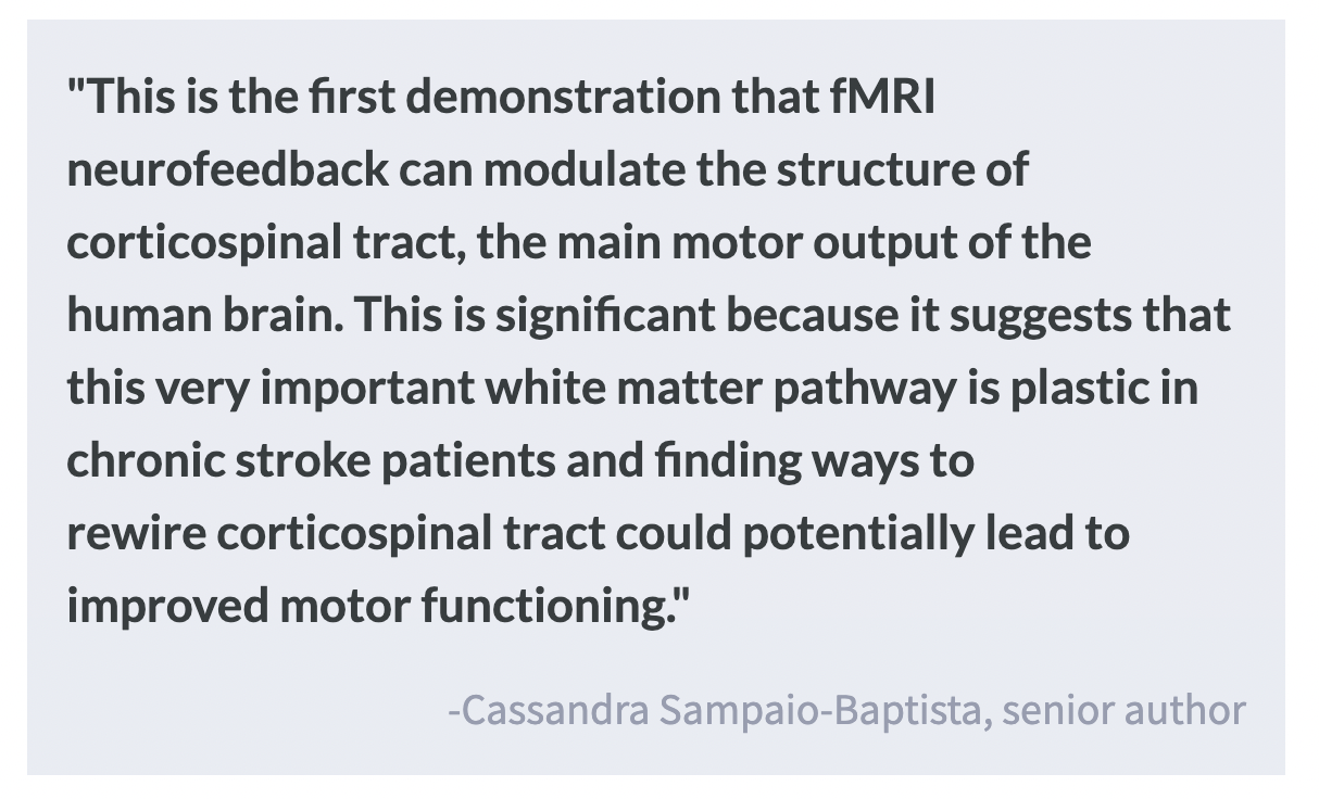
Analysis of the diffusion-weighted data revealed that after 1 week of neurofeedback training, participants experienced a decrease in the asymmetry of white matter structure within the corticospinal tract relative to the sham group. This may reflect an improvement of white matter integrity following neurofeedback, as asymmetry of this white matter bundle has been previously linked with stroke impairment. A correlation between change in white matter structure of the affected corticospinal tract and neurofeedback success was observed in the real group, but not in the sham group. Neurofeedback training did not improve lateralization of brain activity in the visuomotor squeeze task, indicating that there was no carryover effect to other motor functions.
Finally, the authors investigated whether there were changes in brain function outside of the target sensorimotor region and found changes in three significant clusters in the unaffected hemisphere, including the putamen, the lateral occipital cortex, and the parietal operculum cortex, in which the real group increased activity after the neurofeedback training.
What's the impact?
This study finds that stroke survivors were able to use neurofeedback training to increase neural activity in the motor cortex corresponding to their affected limb within a training day, but not across sessions. The neurofeedback also improved gross motor function and reduced structural asymmetry of the corticospinal tract. Overall, this study provides important evidence for the utility of neurofeedback in stroke rehabilitation. Future studies with larger samples and longer follow-ups are needed to determine the utility of this approach for long-term outcomes of stroke patients.