Medial Prefrontal Cortical Neurons Trigger REM Sleep
Post by Shireen Parimoo
The takeaway
The onset of rapid eye movement (REM) sleep – the sleep stage commonly associated with dreaming – was previously associated with activity in subcortical brain regions. Here, the authors show that REM sleep can be regulated by medial prefrontal cortex neurons exerting top-down influence on subcortical areas, thereby triggering REM sleep in mice.
What's the science?
The rapid eye movement (REM) sleep stage is commonly associated with dreaming and is characterized by eye movements as well as an increase in the frequency of theta neural oscillations. Prior work has shown that REM sleep is triggered by subcortical structures like the hypothalamus that in turn give rise to cortical activity. However, it is not known whether cortical neurons can similarly induce REM sleep. This week in Nature Neuroscience, Hong and colleagues used optogenetic stimulation and various imaging techniques to identify the mechanisms by which medial prefrontal and subcortical neurons interact to promote REM sleep.
How did they do it?
The authors injected viral vectors into the medial prefrontal cortex (mPFC) of mice to enhance the expression of light-activated ion channels, which allowed them to stimulate or inhibit the activity of those neurons by shining a laser light. They used two mouse strains; in one strain, they optogenetically manipulated the activity of excitatory pyramidal neurons while in the other strain, they manipulated the activity of inhibitory GABAergic neurons in the mPFC. Optogenetic stimulation was performed under an open-loop setting (i.e., at random times) or under a closed-loop setting (i.e. when the animals entered the REM sleep stage). Open-loop stimulation allowed the authors to observe whether mPFC activity triggered REM sleep from the non-REM (NREM) stage, while the closed-loop stimulation provided insight into the effect of mPFC activation on various features of the REM cycle, respectively.
As the mPFC is widely connected with the rest of the brain, the authors stimulated mPFC neurons terminating in different subcortical regions to identify the subcortical regions that were predominantly activated by mPFC during REM sleep. Next, they used calcium imaging to identify the LH-projecting mPFC neurons according to the sleep stage that they were most active in, that is, during waking, during NREM, and during REM sleep. Of those neurons most active during REM sleep, they further divided them into subgroups based on whether the neurons were relatively more active during wake than during NREM (i.e., R-W-N neurons) or during NREM than during waking (i.e., R-N-W neurons).
Electrodes implanted on the skull were used to obtain recordings of oscillatory brain activity during different sleep stages. Using electroencephalography, the authors recorded changes in power across different frequency bands (e.g., theta and delta), the occurrence of phasic theta events, the duration of REM sleep episodes, and the probability of transitioning between sleep stages (e.g., from NREM to REM sleep). In addition, they performed video-oculography to record eye movement bursts in response to optogenetic manipulation.
What did they find?
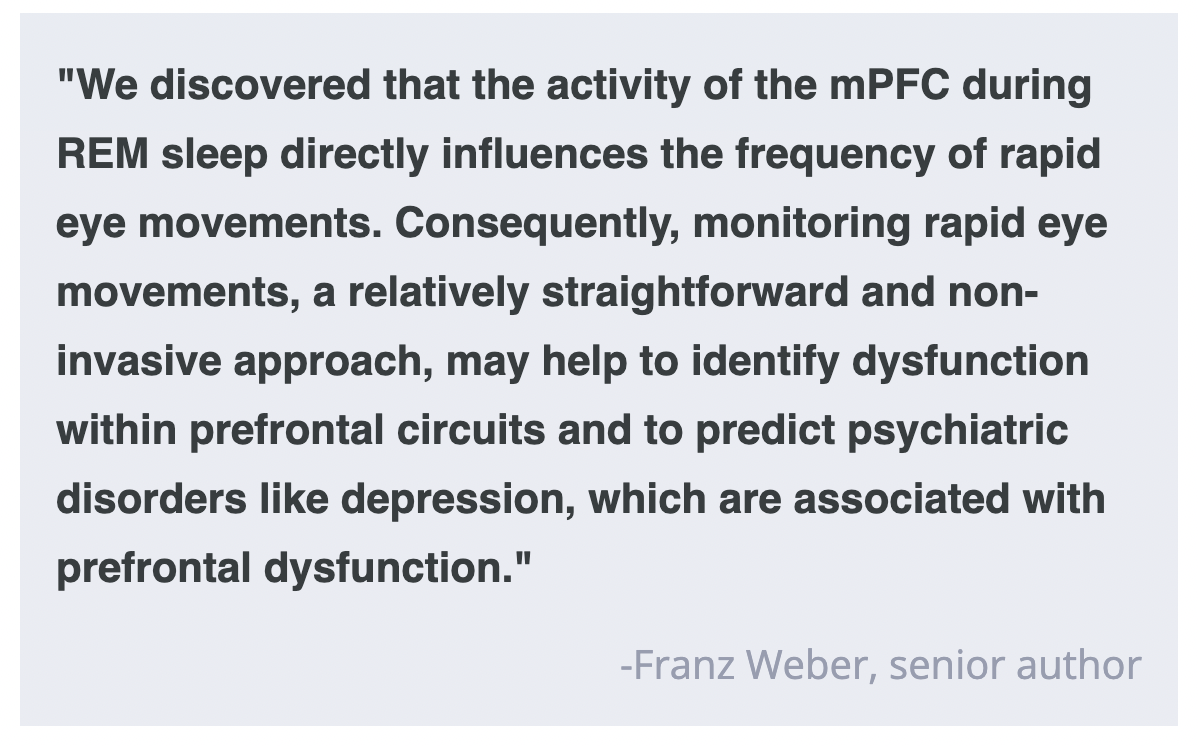
Pyramidal neurons in the mPFC were more active during REM than during NREM sleep and wakefulness. Stimulating pyramidal mPFC neurons increased the transition from NREM to the REM sleep stage, longer REM cycles, and a greater frequency of phasic theta events and eye movement bursts. There was also an increase in theta power with a concomitant reduction in delta power. Inhibition of mPFC pyramidal neurons had the opposite effect, with shorter REM cycles and fewer phasic theta events, along with reductions in theta power and phasic theta events. Conversely, stimulating inhibitory interneurons in the mPFC had a similar effect as inhibiting the pyramidal neurons. These findings indicate that excitatory pyramidal neurons in the mPFC are important for promoting REM sleep.
The authors found that only those neurons projecting from mPFC to the lateral hypothalamus (LH) lateral hypothalamus were involved in REM sleep. Specifically, stimulating LH-projecting pyramidal neurons – but not inhibitory interneurons – led to longer REM sleep episodes, increased theta/sigma power along with reductions in delta power, and an increase in the frequency of phasic theta events and eye movement bursts. Inhibiting these neurons instead reduced REM sleep. Lastly, both the R-W-N and R-N-W subgroups of the LH-projecting neurons were active before the transition to REM sleep, but activity in the R-N-W neurons increased earlier than that in R-W-N neurons and had a faster decline in activity at the end of the REM stage. Moreover, the sustained activity of R-W-N neurons was related to longer REM durations and more theta phasic events, indicating that they may be important for maintaining REM sleep and induced phasic theta events. Altogether, these findings indicate that mPFC pyramidal neurons drive REM sleep through their influence on the lateral hypothalamus.
What's the impact?
This study is the first to demonstrate the mechanisms by which medial prefrontal neurons help induce and maintain REM sleep through their projections to the lateral hypothalamus. These findings provide a more comprehensive understanding of the neuronal circuits underlying REM sleep and may have important implications for treating sleep-related symptoms and disorders.


