Mild Traumatic Brain Injury Increases Risk of Parkinson’s Disease

What's the science?

Mild traumatic brain injury, also referred to as a concussion, is defined as a loss of consciousness or confusion due to head injury that lasts less than 30 minutes. Mild traumatic brain injury is especially common among the elderly, athletes and in the military, and is increasingly associated with risk for psychiatric and neurodegenerative diseases. The relationship between mild traumatic brain injury and risk for Parkinson’s disease is not well understood. This week in Neurology, Gardner and colleagues assess the risk of Parkinson’s disease after mild traumatic brain injury in a large sample of patients.

How did they do it?

Participants (18 years or older) from the Veterans Health Association (military veterans) with a diagnosis of traumatic brain injury were included and were age matched to a random sample of participants without brain injury (325,870 total participants). They diagnosed Traumatic brain injury using detailed clinical assessments or criteria from the Department of Defense (IC-9 codes). They also assessed other diseases at baseline and Parkinson’s disease at least one year after baseline in this longitudinal study. They then used a Cox Proportional Hazards model (a standard statistical model for assessing longitudinal risk) to determine risk of developing Parkinson’s disease with and without traumatic brain injury, including assessment of risk specifically after mild traumatic brain injury. They controlled for confounding variables including medical conditions, psychiatric disease and demographics: age, sex, ethnicity, education and level of income.

What did they find?

A total of 1462 participants developed Parkinson’s disease over the course of the study (average 4.6 years follow-up), 65% of whom had previously experienced traumatic brain injury. Participants with previous traumatic brain injury were significantly more likely to develop Parkinson’s disease than those without traumatic brain injury (hazards ratio: 1.71). Even mild traumatic brain injury was associated with increased risk (56%) of developing Parkinson’s disease after adjusting for all confounding variables.

Relationship between Traumatic Brain Injury and Parkinson’s disease

What's the impact?

This is the first large-scale, nationwide study to demonstrate that mild traumatic brain injury is associated with an increased risk of Parkinson’s disease. We now know that focusing on the prevention of mild traumatic brain injury will be important for reducing risk of Parkinson’s disease.

itbiquote.png

R. Gardner et al., Mild TBI and risk of Parkinson disease: A Chronic Effects of Neurotrauma Consortium Study. Neurology (2018). Access the original scientific publication here.

How Deep Brain Stimulation Affects Decision-Making in Parkinson’s disease

What's the science?

How much time should we give ourselves to make a decision? For example, when faced with a difficult decision, we might give ourselves more time to garner more evidence before we reach the ‘decision threshold’ and decide. One brain region involved in adjusting our decision threshold (meaning we take more or less time before the decision) is the subthalamic nucleus (STN). Deep brain stimulation (DBS) of the STN is often performed to reduce motor symptoms in Parkinson’s disease, however, a negative side effect can be impairment in adjusting the decision threshold, leading to impulsive responses. This week in Current Biology, Herz and colleagues conducted a study in patients with DBS electrodes for Parkinson’s placed in the STN, in order to assess how stimulation of this site affects the decision threshold.

How did they do it?

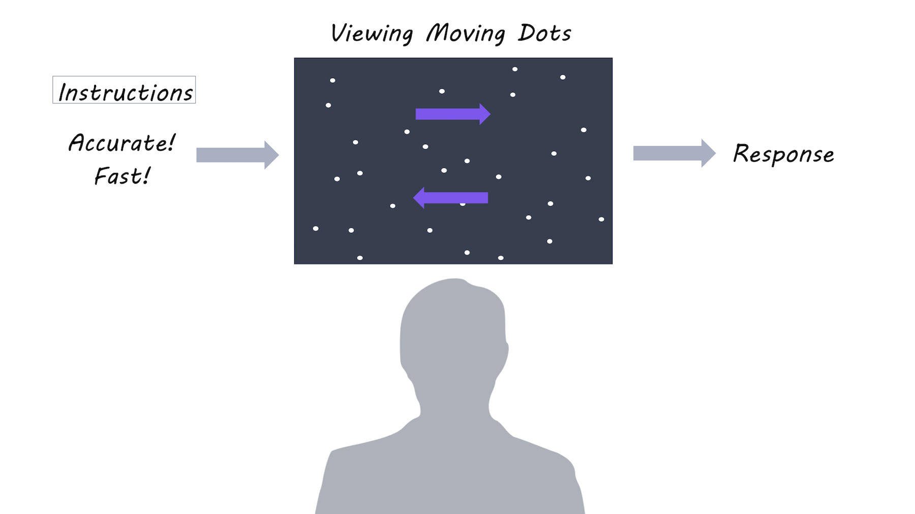
Ten patients with Parkinson’s participated in the study after undergoing surgery to implant electrodes for DBS in the STN. Each patient performed a decision-making task: 1) when DBS was off 2) with DBS on continuously and 3) with ‘adaptive DBS’ where DBS only turns on when necessary. The decision-making task involved looking at dots moving on a screen, and deciding whether the majority of dots were moving to the left or to the right. There were two task conditions and two forms of instruction: In the easy condition, 50% of the dots moved in the same direction, while in the difficult condition only 8% of dots moved in the same direction. Participants were also instructed to focus on either speed or accuracy of their decision.

Dot motion perception

What did they find?

When DBS was off, participants responded more slowly during the difficult task and when instructed to focus on accuracy (versus speed). However, when DBS was on, slowing during a difficult task was diminished, but slowing due to focus on accuracy remained the same. During adaptive DBS, stimulation came on at different times across trials (when beta activity happened to be high). When the DBS stimulation came on during a 400-500 ms time window after the moving dots appeared on the screen, the time required to make a decision (usually increased during the difficult task) was most diminished, suggesting that the effect of stimulation is confined to a short time window. Using ‘drift diffusion modelling’, they found that stimulation affected the decision threshold time specifically, as opposed to, for example, the motor response time. While DBS was off, beta activity increased after presentation of the dots during the difficult condition, and was related to the decision threshold, but these effects were abolished during stimulation. This indicates that DBS may be lowering the decision threshold by changing the relationship between STN activity and threshold adjustments.

What's the impact?

These results are the first to show that the STN may be directly involved in decision thresholds (how much evidence we need before we reach a decision). During a narrow time window, the STN adjusts decision thresholds based on the anticipated difficulty of the decision. This may be a mechanism by which decision-making is impaired in people with Parkinson’s who have DBS.

brown_quote_week11_small.png

P. Brown et al., Mechanisms Underlying Decision-Making as Revealed by Deep-Brain Stimulation in Patients with Parkinson’s Disease. Current Biology (2018). Access the original scientific publication here.


Dyskinesias in Parkinson’s disease are Caused by a Subgroup of Neurons

What's the science?

In Parkinson’s disease, dopamine neurons in the midbrain degenerate resulting in problems with body movement. A dopamine medication called levodopa can be very effective for improving symptoms, however, in some cases it causes involuntary movements called dyskinesias. We know that unwanted neural activity in brain regions such as the striatum, motor cortex and sensorimotor cortex may be involved, but the specific brain region and cells causing dyskinesias are not known. Recently in Neuron, Girasole and colleagues identify a subgroup of neurons responsible for dyskinesias.

How did they do it?

They first used a method called Targeted Recombination in Active Populations (TRAP) in transgenic (genetically modified) mice. TRAP allows certain proteins (acting as labels) to be expressed in active neurons (as opposed to inactive neurons). In mice with levodopa-induced dyskinesias, they identified neurons that were active during the dyskinesias compared to control mice. Second, they then used optogenetics: Controlling neuron activation by shining light on genetically modified neurons of interest. This allowed them to inhibit and activate these specific neurons in the mice to see if they played a causal role in dyskinesias.

What did they find?

Only neurons in the striatum were significantly more active during dyskinesias compared to control mice. When examining these neurons more closely, they found that most of the active neurons were medium spiny neurons (a specific cell type of neuron found in the striatum) that were part of the 'direct pathway', an inhibitory pathway involved in motor function that is defective in Parkinson’s disease. When these neurons were inhibited with optogenetics, the dyskinesias were reduced. Inhibiting the activity of neurons in the motor or sensorimotor cortices did not reduce dyskinesias, demonstrating a causal role for striatal neurons in producing medication-induced dyskinesias.

Images are generated by Life Science Databases(LSDB)., Striatum, colour by BrainPost, CC BY-SA 1.0

Images are generated by Life Science Databases(LSDB)., Striatum, colour by BrainPost, CC BY-SA 1.0

What's the impact?

This is the first study to identify the neurons within the striatum that cause dyskinesias in mice. Dyskinesias are a detrimental side effect of levodopa in Parkinson’s disease and can be debilitating to patients who experience them. Understanding which neurons cause dyskinesias brings us one step closer to finding a way to treat them.

Reach out to study author Ally Girasole on Twitter @AllyGirasole

A. E. Girasole et al., A Subpopulation of Striatal Neurons Mediates Levodopa-Induced Dyskinesia. Neuron. 97, 1–9 (2018). Access the original scientific publication here.