Ripples During Slow-Wave Sleep Re-balance Neurons

What's the science?

Synapses in the brain are strengthened while awake and synaptic depression (weakening of the synapses) occurs during slow-wave sleep to rebalance synapses. This synaptic depression may help to selectively preserve memories, however, how it occurs during sleep is unclear. During slow-wave sleep, the hippocampus emits high frequency oscillations called “sharp-wave ripples” which reactivate the neurons involved in recent memories. Ripples could be required for synaptic depression during sleep, making “room” for new memories to form. This week in Science, Norimoto and colleagues test whether ripples are required for synaptic depression and subsequent memory formation in mice.

How did they do it?

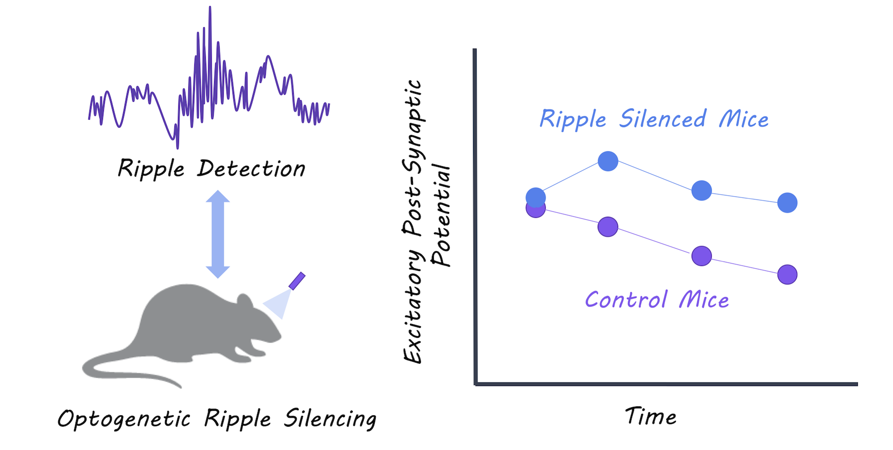
They silenced sharp-wave ripple events during slow-wave sleep using optogenetic-feedback (i.e. every time a ripple event was detected, they inhibited the hippocampal neurons to reduce firing) in a group of test mice. They then recorded excitatory postsynaptic potentials (activity in the postsynaptic neurons) during slow-wave sleep and compared the test mice to control mice to determine whether synaptic depression during sleep was affected in the test mice. Mice then underwent a spatial object-recognition task. First, they explored an area containing two new objects, and then returned to the same location 2 hours later where one of the objects had moved. The authors compared the memory performance between test and control mice to determine whether disrupting the ripples had an effect on new memory acquisition.

What did they find?

The authors were able to successfully reduce almost all of the ripple events in the test mice. Control mice experienced depressed postsynaptic activity representing the expected synaptic depression that occurs during slow-wave sleep, while mice with the impaired ripple events did not show synaptic depression. Control mice showed normal memory performance, while mice with impaired ripple events were unable to identify a moved object in the spatial object recognition task, suggesting that they could not form new memories. The authors were also able to replicate the lack of synaptic depression by testing the effects of disrupting ripples in hippocampal tissue slices. They used in vitro and in vivo experiments to show that synaptic depression occurring during slow-wave sleep is mediated by NMDA receptors.

Optogenetic ripple silencing

What's the impact?

This is the first study to link sharp-wave ripples during slow-wave sleep with synaptic depression and memory performance. Before this study, we did not understand the mechanism through which synaptic depression occurred during slow-wave sleep. We now know that ripples during slow-wave sleep are critical for balancing of synapses and for new memory formation.

Yuji_quote_week11_small.png

H. Norimoto et al., Hippocampal ripples down-regulate synapses. Science (2018). Access the original scientific publication here.

 

A Model for the Spread of Tau through Connected Tracts in the Human Brain

What's the science?

In Alzheimer’s disease, tau proteins accumulate in the hippocampus resulting in neurofibrillary tangles. Beta-amyloid plaques, another form of protein aggregation, are thought to help tau proteins spread. One way that tau may spread from neuron to neuron is through neural connections, while another possibility is that it simply spreads to neurons located close by. This week in Nature Neuroscience, Jacobs and colleagues used brain imaging to ask: ‘How does  tau spread?’

How did they do it?

Healthy older participants from the Harvard Aging and Brain Study were scanned over several years with positron emission tomography (PET) imaging to measure tau and beta-amyloid in the brain, and diffusion tensor imaging (DTI) to measure connectivity (of white matter tracts) in the brain. They tested whether beta-amyloid in the brain at baseline predicts hippocampal volume loss. They then measured whether this volume loss predicts abnormalities in the hippocampal cingulum bundle (a white matter tract that innervates the hippocampus and connects it with the posterior cingulate cortex) and in turn, whether these abnormal connections predict the accumulation of tau in the posterior cingulate cortex. They ran control analyses with another tract (that does not innervate the hippocampus) and another close by region. Associations with memory and executive functions were also assessed to understand the clinical relevance. 

What did they find?

Brain beta-amyloid level at baseline predicted hippocampal volume loss. The hippocampal volume loss also predicted abnormal white matter tract connectivity over time in the hippocampal cingulum bundle, but not in other white matter tracts close by that do not directly connect with the hippocampus. The abnormal connectivity in this tract predicted the accumulation of tau in a connected region called the posterior cingulate cortex, but not in another adjacent control region. Collectively, these changes were associated with memory decline over time. This means that early Alzheimer’s pathology (beta-amyloid) initiates a cascade of hippocampal volume loss followed by abnormal tract connectivity and the spreading of tau along this tract. 

Spread of tau and beta-amyloid accumulation over time

What's the impact?

This is the first study to confirm that tau likely spreads via neural connections (rather than just to regions close by) from the hippocampus, facilitated by beta-amyloid in the brain. Clarifying the order in which Alzheimer’s pathology spreads, as well as the mechanism through which it spreads is critical for helping to target the advancement of Alzheimer’s disease.

DrJacobs_small.jpg

You can reach out to her about her work at @DrHeidiJacobs on Twitter.

H. I. L. Jacobs et al., Structural tract alterations predict down-stream tau accumulation in amyloid positive older individuals. Nat. Neurosci. (2018). Access the original scientific publication here.

The Link Between a High Salt Diet and Impaired Brain Function in Mice

What's the science?

A diet high in salt has been linked to altered brain blood supply, stroke and cognitive impairment. Evidence suggests that the immune system becomes activated in response to salt, and may be involved. Recently in Nature NeuroscienceFaraco and colleagues report a mechanism linking salt in the gut to immune system activation and reduced blood flow in the brain.

How did they do it?

They fed mice high salt diets and measured immune system markers in the blood and the function of cells lining brain blood vessels (endothelial cells which work to allow blood flow) at set time intervals over a 24 week period. They also measured resting brain blood flow using magnetic resonance imaging (MRI) with arterial spin labelling.

What did they find?

Mice fed a high salt diet had lower brain blood flow and dysfunctional endothelial cells. Additionally, they had worsened memory function, and a reduced ability to perform daily activities. Mice fed a high salt diet also had higher levels of immune cells (lymphocytes) in the gut, which resulted in higher inflammatory markers (IL-17) in the blood. Importantly, the lowered brain blood flow and cognitive problems were dependent on this immune system response.

Relationship between the gut and the brain

What's the impact?

This study confirms previous theories that the immune system plays a role in linking dietary salt with brain function. Importantly, it reveals a specific immune system pathway linking the gut and the brain. This gut-brain pathway could be targeted with therapies to prevent harmful effects of salt on the brain. 

G. Faraco et al., Dietary salt promotes neurovascular and cognitive dysfunction through a gut-initiated TH17 response. Nat. Neurosci. (2018)

Access the original scientific publication here.