Understanding Compulsive Disorders

Post by Laura Maile

What is compulsion?

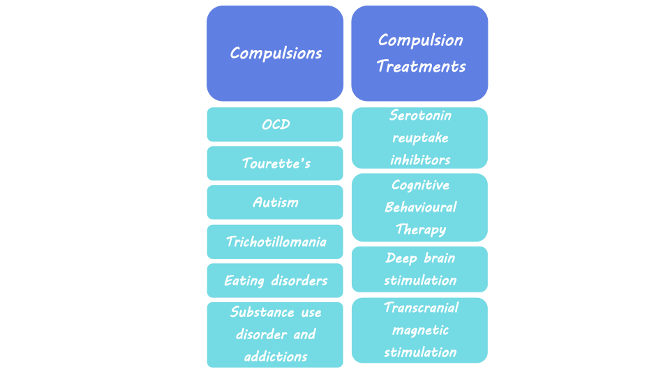
Compulsions are described as persistent urges to perform a specific behavior, even when the consequences are negative. They induce a feeling that the individual must complete the behavior, even though it may conflict with their overall goal. These repetitive behaviors are maladaptive, meaning they prevent individuals from adapting to their environment and are detrimental to overall well-being. Compulsions are a symptom of diseases like Obsessive Compulsive Disorder (OCD) and Tourette’s Syndrome, but they can occur in many other psychiatric disorders including Autism Spectrum Disorder, trichotillomania, eating disorders, substance use disorder, and other addictions. While compulsivity is a maladaptive symptom of disease, it can also be a component of everyday human behavior that leads to positive outcomes, including behaviors such as proofreading or athletic performance rituals.  

Neurological basis of compulsive behavior

Some theories of compulsive behavior, which generally include ideas of positive and negative reinforcement learning, suggest that the neural networks that control goal-directed behavior and habits are out of balance. These networks include brain regions important for goal-directed and habitual behavior like the striatum, a part of the basal ganglia, which is important for controlling motor behavior, learning, and decision-making. In rodent studies where drugs are administered to inactivate specific areas of the striatum, habitual seeking of food and alcohol is reduced. Rodent studies also show that compulsive behavior can be born out of negative reinforcement, or avoidance of negative outcomes. The emergence of this compulsivity is dependent on brain systems required for habit formation like the striatum. The medial prefrontal cortex (mPFC) is also important for goal-directed behavior, habit formation, behavioral flexibility, the association between actions and outcomes, and decision-making. The nearby orbitofrontal cortex (OFC), a region involved in decision-making related to reward and emotion, is also likely involved in compulsive behavior. Another region implicated in goal-seeking and habitual behavior is the amygdala, which is part of the limbic system associated with emotion, fear, and aspects of learning and memory that are influenced by emotionally important stimuli.  

Compulsive behavior can result when there is an imbalance or impairment in these networks that regulate goal-directed behavior. Neuroimaging studies in the human brain suggest that changes in these brain regions that regulate behavior are related to compulsion. For example, studies show reduced brain volume (i.e., grey matter) and elevated brain activity in the OFC in individuals with OCD. Changes in neural connectivity between the prefrontal cortex and striatum are also seen in patients with OCD.  

Human imaging studies of individuals with OCD and substance use disorder found reduced functional connectivity (i.e., neural synchrony between brain regions) and activity of prefrontal cortex regions, which correlated with compulsive behavior. This loss of connectivity in the regions governing goal-directed behavior suggests a loss of control over habitual behavior and an inability to inhibit future actions, leading to compulsivity. Changes in activity related to compulsivity have also been seen in the insula, a region of the cortex associated with interoception, or awareness of bodily feelings. Interoceptive feelings and insula activity may trigger habits that have been developed through experiences involving negative reinforcement. The variety of findings in human imaging studies demonstrates the complexity of these neural networks that interact to influence compulsive behavior and highlight the need for more detailed research.

Treating compulsive behavior

Treatments for OCD and compulsive behaviors associated with other psychiatric disorders include serotonin reuptake inhibitors, cognitive behavioral therapy, and deep brain stimulation.  Deep brain stimulation includes the implantation of electrodes into the brain to allow for electrical stimulation of specific areas. Clinical studies have shown that deep brain stimulation of the nucleus accumbens, a region associated with reward, was successful at reducing compulsivity symptoms in OCD patients who did not respond to other forms of treatment. A less invasive form of treatment is transcranial magnetic stimulation, which uses a magnetic coil outside the skull to influence the activity of certain brain areas. Transcranial magnetic stimulation of the mPFC has been shown to reduce OCD symptoms in some patients.  Other studies using this technology to stimulate regions like the OFC, striatum, and basal ganglia show some promise for treating intractable OCD, though results are mixed.  Transcranial magnetic stimulation has also been successfully deployed to target regions of the prefrontal cortex to reduce compulsive drug seeking in patients with substance use disorder.  

What's next?

While some success has been achieved with transcranial and deep brain stimulation, more research is needed to improve outcomes and reduce side effects of compulsive behavior. New deep brain stimulation techniques are being investigated that can deliver neurostimulation while recording neural activity, which will allow scientists more insight into the neural basis of compulsive behavior. Other novel treatments being studied for compulsive behavior include ketamine and psychedelics, which can be used as treatments for depression or anxiety. There is also ongoing work aimed at uncovering how findings related to the contribution of serotonin, dopamine, and noradrenaline to compulsive behavior translate from rodent to human systems.  

References +

Robbins et al. From compulsivity to compulsion: the neural basis of compulsive disorders. Nat. Rev. Neurosci. (2024).

Robbins et al. Obsessive-Compulsive Disorder: Puzzles and Prospects. Neuron (2019).

Marti-Pratz et al. The development of compulsive coping behavior depends on dorsolateral striatum dopamine-dependent mechanisms. Nature Portfolio (2023).

Simmler et al. Neural circuits in goal-directed and habitual behavior: Implications for circuit dysfunction in obsessive-compulsive disorder. Neurochemistry International (2019).

Anticevic et al. Global resting-state functional magnetic resonance imaging analysis identifies frontal cortex, striatal, and cerebellar dysconnectivity in obsessive-compulsive disorder. Biol. Psychiatry (2014).

Denys et al. Deep Brain Stimulation of the Nucleus Accumbens for Treatment-Refractory Obsessive-Compulsive Disorder. JAMA Psychiatry (2010).

Goodman et al. Harmonizing the Neurobiology and Treatment of Obsessive-Compulsive Disorder. American Journal of Psychiatry (2021).

How Physical Activity Alters the Brain’s Stress Pathways and Cardiovascular Disease Risk

Post by Lani Cupo

The takeaway

One of the ways that physical activity reduces the risk of cardiovascular disease is by reducing activity in the brain’s stress network.   

What's the science?

Scientific evidence confirms what many people feel; physical activity (especially cardio) is good for your heart and also helps manage stress levels. The mechanisms underlying these benefits, however, are still poorly understood, and so is the degree to which stress reduction contributes to the cardiovascular benefits of exercise. This week in the Journal of the American College of Cardiology, Zureigat and colleagues investigated the underlying mechanisms linking physical activity, stress circuits, and cardiovascular health.

How did they do it?

The authors studied about 50,000 adults enrolled in a Biobank study who filled out a health behavior survey that included details on their physical activity. Information on cardiovascular events (such as heart failure or stroke) and psychiatric disorders (such as depression) before and after enrolment were derived from the electronic medical records. A subset of 744 participants also underwent brain imaging with F-FDG positron emission tomography (PET)/computed tomography (CT), which provides a marker for regions with high glucose metabolism as a proxy for brain activity. The authors calculated the ratio of PET signal in the amygdala and the ventromedial prefrontal cortex (referred to as AmygAc), where a higher value indicates more stress-related activity. They chose to study these regions because they have previously been associated with chronic stress and related syndromes.

The health questionnaires provided information on the history of cardiovascular events (such as heart failure or stroke) and psychiatric disorders (such as depression). The authors used regressions to statistically assess relationships among the variables. First, they examined the associations between physical activity and AmygAc. Then they assessed the association between physical activity and cardiovascular events. Finally, they assessed the association between AmygAc and cardiovascular events. With these sets of equations, they were able to assess whether AmygAc mediated the relationship between physical activity and cardiovascular events. That is to say, whether physical activity indirectly impacts cardiovascular health via stress reduction in the brain’s stress pathways. History of depression was also included as an interaction variable in these models to allow the authors to understand whether patterns in the relationships among the other variables were different for people with past depression.

What did they find?

First, the authors found that physical activity was associated with a decrease in stress-related neural activity, such that the more physical activity participants reported, the less stress-related activity they showed. This was, in large part, associated with increases in prefrontal cortical activity, which may contribute to cognitive health benefits. Second, as expected, they found that physical activity was associated with decreased risk of cardiovascular events. Third, they found that increased AmygAc, representing increased stress-related activity, was associated with an increased risk of cardiovascular events. These three findings aligned well with their hypotheses: 1) more physical activity is related to less stress activity in the brain, 2) more physical activity is related to less risk of cardiovascular events, and 3) more stress-related activity is related to more risk of cardiovascular events.

Next, they evaluated whether physical activity lowers cardiovascular disease risk by reducing AmygAc. From their mediation analysis, the authors found that AmygAc was a partial mediator of the relationship between physical activity and cardiovascular events. This means that one way in which physical activity is associated with improved cardiovascular health is by reducing stress, but it’s not the only underlying mechanism.

Finally, the authors found that the benefits of physical activity on cardiovascular health were highest among people with a history of depression. Specifically, people without a history of depression experienced the anticipated benefits of physical exercise, and their risk reductions plateaued at ~300 minutes of moderate-intensity activity per week. On the other hand, people with a history of depression derived roughly double the overall cardiovascular risk reductions compared to those without depression, and continued to derive benefits at the higher levels of activity.

What's the impact?

In this paper, the authors demonstrate that physical activity reduces stress-related brain activity and the risk of cardiovascular events, especially for people with a history of depression. Importantly, reduced stress partially mediates the relationship between physical activity and cardiovascular events, providing an indication of the underlying mechanisms. Future work may further characterize other mediating factors among these variables.

A Newly Identified Body-to-Brain Circuit for Regulating Inflammation

Post by Shahin Khodaei

The takeaway

Communication between the brain and the body plays a key role in the immune response: information about bodily inflammation is delivered to the brain, which can subsequently adjust inflammation in the body to an appropriate level.

What's the science?

Inflammation, a component of innate immunity, is an important part of the body’s response to damage or pathogens. Several studies have shown that the brain can detect the state of inflammation in the body and that neurons can modulate inflammation through their activity. However, the pathways by which the brain regulates bodily inflammation are still not well understood. This week in Nature, Jin and colleagues published a study that identified: 1) specific populations of neurons that carry information about the state of inflammation in the body to the brain, and 2) specific populations of neurons in the brain that respond to this information to ramp up or dampen inflammation in the body.

How did they do it?

To cause inflammation in mice, the authors injected them with lipopolysaccharide (LPS) – a component of bacterial cell walls that is recognized by immune cells as a signal for pathogens. In response, these cells release protein messengers known as cytokines, which can be pro-inflammatory (heighten inflammation) or anti-inflammatory (reduce inflammation). 

The authors then had two major goals: to determine which neurons respond to inflammation and to determine how the activity of these neurons shapes the inflammatory response.

For their first goal, the authors used imaging techniques to find out which neurons are activated after LPS, and pro- or anti-inflammatory cytokines. They then used single-cell RNA sequencing to map the genes that are expressed by these neurons, to find markers to identify them.

To manipulate the activity of neurons, the authors used the genetic technology of designer receptors exclusively activated by designer drugs or DREADDs. Depending on the type of DREADD (excitatory vs. inhibitory) inserted into cells, the authors can selectively increase or decrease the activity of sub-populations of neurons. In conjunction, the authors used another genetic technology called TRAP (targeted recombination in active populations), which allowed them to express DREADDs only in neurons that were involved in inflammation. This approach lets them manipulate the activity of inflammation-responsive neurons.

What did they find?

Using their imaging techniques, the authors found a population of neurons in the brainstem that was activated by LPS-triggered inflammation. These neurons received information about bodily inflammation from the vagus nerve. The authors found two distinct populations of vagal neurons: one responding to pro-inflammatory cytokines and expressing the genetic marker TRPA1, and the other to anti-inflammatory cytokines and expressing the genetic marker CALCA. In this way, vagal neurons transmit information regarding the inflammatory state to the brainstem.

How does manipulating the activity of these neurons affect the inflammatory response? Using DREADDs, increasing the activity of brainstem neurons was anti-inflammatory, while inhibiting their activity during LPS injection led to much higher inflammation. Similarly in the vagus nerve, increasing the activity of either the TRPA1 or the CALCA subpopulations had an anti-inflammatory effect. Remarkably, the authors further showed that in a mouse model of deadly inflammation, activating this body-brain circuit using DREADDs significantly reduced the likelihood of death.

What's the impact?

This study identified a specific body-to-brain circuit that regulates inflammation. By monitoring the levels of pro- and anti-inflammatory cytokines in the body, this circuit monitors the inflammatory response and regulates inflammation levels as needed. In the future, targeting this system may provide new strategies for treating diseases that involve dysregulation of the inflammatory response.

Access the original scientific publication here.