Identifying an fMRI Biomarker for Cognitive Decline in Alzheimer’s Disease

Post by Kelly Kadlec

The takeaway

Two fMRI-based metrics previously used to evaluate cognitive decline with age may also be useful for assessing both the risk and severity of Alzheimer’s disease. These scores can help distinguish between rates of memory decline in healthy individuals and those with varying levels of risk for developing Alzheimer’s.

What's the science?

A formal diagnosis of Alzheimer’s disease (AD) is often preceded by progressive stages of cognitive decline. At each of these stages, patients are at varying risk for advancing to AD, but assessing the risk of an individual is difficult due to a high degree of heterogeneity in neurocognitive aging. Previously, functional magnetic resonance imaging (fMRI) contrast maps for novelty and memory tasks have yielded two corresponding single-value scores that have been proposed as biomarkers of neurocognitive aging. This week in Brain, Soch and colleagues compare these fMRI-based scores in healthy individuals, individuals with AD, and individuals in different risk categories for developing AD, to assess their ability to distinguish between clinical and healthy rates of cognitive decline.

How did they do it?

This study comprised five groups of individuals: healthy controls with no family history of AD, healthy individuals with a first-degree relative with AD, patients with AD, and patients in one of two symptom-based risk states for AD: mild cognitive impairment (MCI) or subjective cognitive decline (SCD), where MCI is considered the more severe.

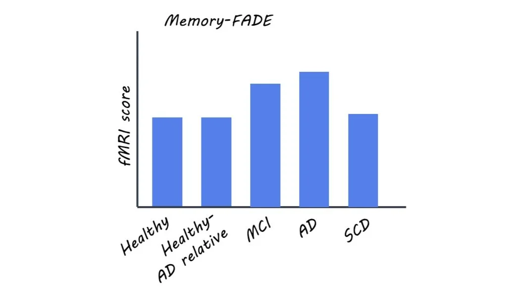
The authors collected fMRI data from the participants during image-based novelty and memory tasks. They used the resulting contrast maps to calculate two scores: Functional Activity Deviation during Encoding (FADE) and Similarity of Activations during Memory Encoding (SAME). Additionally, psychometric and genetic testing was done for each participant, and a subset had amyloid positivity testing. The authors hypothesized that increasing FADE scores and decreasing SAME scores would be associated with worse AD severity and higher risk for AD.

What did they find?

The authors found that memory and novelty-based FADE and SAME scores could be used to distinguish between the different participant groups and also correlated with known risk factors and cognitive assessments.

The authors reported that increasing risk for AD corresponded to larger deviations in FADE and SAME scores (i.e. more atypical fMRI results). Only memory-based FADE and SAME scores differentiated between the two more severe clinical groups (AD and MCI) and all other participant groups, and only novelty-based scores distinguished between AD and MCI patients.

The authors confirmed that FADE and SAME scores for memory and novelty tasks corresponded to other currently used psychometric tests of cognitive decline and AD severity. The authors also found that within the AD-related participants, higher FADE and lower SAME scores corresponded to the presence of an AD genotype. In addition, the authors found that especially novelty-based scores were sensitive to amyloid positivity.

To demonstrate the potential clinical value of these fMRI biomarkers, the authors used FADE and SAME scores to predict diagnostic groups for the participants and classified each group with above-chance accuracy. They also used these scores to predict the presence of an AD genotype in participants with AD relatives.

What's the impact?

Proper assessment of AD risk and severity is challenging, and this study proposes two promising neural biomarkers. These fMRI-based scores distinguished between differing stages of the disease and predicted other proposed risk factors for AD. This knowledge is critical for choosing the correct treatment routes and improving diagnosis accuracy earlier in the development of AD. Further, a longitudinal study is needed to determine how predictive they are of future outcomes (i.e. MCI progressing to AD). 

Access the original scientific publication here.

How Brain Connectivity Contributes to Different Types of Goal Pursuit

Post by Lila Metko

The takeaway

There is lower connectivity between movement-related brain regions in individuals with a higher propensity to use the ‘prevention system’ to pursue goals that prevent a negative outcome rather than the ‘promotion system’ to pursue goals that have a positive outcome.

What's the science?

According to regulatory focus theory, there are two major cognitive-motivational systems involved in accomplishing goals: the promotion system, involved in achieving hopes and dreams, and the prevention system, involved in fulfilling duties and obligations. The promotion system is oriented towards making good things happen, while the prevention system is focused on preventing negative things from happening. Individual differences in these systems of self-regulation have been linked to psychopathology. There are previously established brain regions that have been shown to be involved in each of these systems and they share some overlap. This week in PNAS Nexus, Kim and colleagues used functional magnetic resonance imaging (fMRI) data to create a predictive network model capable of predicting differences in propensity towards regulatory focus systems based on connectivity between neural structures. 

How did they do it?

The authors studied 1,307 university students enrolled in the Duke Neurogenetics Study. They gave the participants the Adolescent Regulatory Focus Questionnaire (RFQ), a questionnaire that measured their inclination towards a promotion system or prevention system way of attaining goals. fMRI scans were taken of the participants in both resting state (patients awake with eyes open but with no task) and during emotional face matching, card guessing, working memory, and face naming tasks. The authors then estimated general functional connectivity for each participant by combining resting state and task fMRI data and regressing out task-related events, to obtain more reliable data than if they used resting fMRI alone. A predictive model of regulatory focus orientation was created using correlations from participants’ functional connectivity to their scores in the RFQ. A group of participants was left out of the creation of the model to test its ability to predict regulatory focus orientation. To test this, they compared actual scores to predicted scores in the group that was left out. 

What did they find?

The model generated by the authors was predictive of prevention but not promotion scores. It’s possible the lack of prediction for promotion scores is because promotion-orientated processes do not require as complex cognitive processes as prevention and thus may not be detectable in these measures of functional connectivity. It was found that lower functional connectivity between association cortices (brain regions involved in understanding sensory information and planning a behavioral response) was correlated with increased prevention. It had been previously established that these association regions were important for prevention behaviors, but interestingly the authors found a novel region associated with prevention; the primary motor cortex. More than half of the measures of brain connectivity that had a negative correlation with prevention scores involved the primary motor cortex.  

What's the impact?

This study is the first to show that the primary motor cortex, a region involved in initiating voluntary movements, contributes to prevention system function. It is also the first study to create a predictive model for these types of preventative behaviors in a large sample. Importantly, regulatory focus orientations are predictive of vulnerability to psychopathology and improper function of these regulatory systems may also be predictive of generalized anxiety disorder and depression. Thus, the study of brain regions involved in regulatory focus is of high clinical significance.  

Access the original scientific publication here.

REM Sleep Protects Against Fearful Memories

Post by Meagan Marks

The takeaway

REM sleep may protect against the development of post-traumatic stress disorder by enhancing the brain’s ability to extinguish fearful memories. It does so by strengthening the excitability of infralimbic cortex neurons, which play a crucial role in fear extinction. 

What's the science?

Post-traumatic stress disorder (PTSD) often presents as persistent, uncontrollable fear responses triggered by cues associated with a past traumatic event. PTSD is likely caused by a neural disruption in fear extinction, which is the process of learning that a fear-inducing cue is no longer predictive of danger. Fear extinction is carried out by neurons in the infralimbic cortex (IL) in mice, which is homologous to the ventromedial prefrontal cortex in humans. It is a region highly active during rapid eye movement (REM) sleep. Interestingly, PTSD patients often experience disturbances in REM sleep, but the exact role that it plays in fear extinction remains unknown. This week in Current Biology, Hong and colleagues determine how REM sleep influences fear extinction, particularly through its influence over IL neuron excitability.

How did they do it?

To understand the paired role that REM sleep and IL neurons play in fear extinction, the authors conducted an experiment over the course of three days. On the first day, the mice were fear conditioned, learning that an auditory cue (20-second tone) would elicit an unpleasant stimulus (one-second foot shock). Immediately after this conditioning, the authors used optogenetics to silence the activity of IL neurons during sleep. At this stage, the mice were split into three groups: for one group, IL neurons were silenced only during REM sleep, while for another, the neurons were silenced immediately after REM sleep. A control group with no neural manipulation was also included. 

On the second day, the mice were placed back in the fear conditioning environment, but this time, the auditory cue was played without a subsequent foot shock. This was a day of extinction learning, where the mice learned that the auditory cue should no longer induce fear. 

On the third and final day, the auditory cue again played with no subsequent foot shock. This was a day of recall, where the authors observed how well the mice extinguished the fear association from the day before. They did so by measuring how long the mice froze after each auditory cue, an innate fear response in rodents. 

Additionally, the authors repeated this paradigm twice more, with slight changes. During one repeat, the authors waited an additional four hours after conditioning to silence IL neurons during REM sleep. During the second, IL neurons were not silenced after fear conditioning but instead were immediately silenced after extinction learning.

What did they find?

The authors found that inhibiting IL activity during REM sleep post-conditioning significantly increased freezing time during recall. The control mice and mice with inhibition after REM sleep did not freeze as much, suggesting that IL activity during REM sleep is crucial to the consolidation of fear extinction memories. Delaying the post-conditioning inhibition of IL activity during REM sleep did not significantly alter freezing, nor did it inhibit IL neurons immediately after extinction learning.

Looking at these findings altogether, it can be concluded that IL neuron activity during REM sleep that occurs immediately after fear conditioning is crucial to fear extinction. This is because inhibiting IL neuron activity decreases the overall excitability of the neurons, and therefore, inhibiting IL neuron activity during all of REM sleep when the neurons are most active decreases their excitability the following day. This hindered their ability to fire during extinction learning, and the mice were less capable of encoding new fear extinction memories. 

What's the impact?

This study found that REM sleep plays a crucial role in fear extinction by strengthening the excitability of IL neurons and enhancing their ability to encode new extinction memories. This is an important finding when it comes to PTSD, as many patients struggle with disturbances in REM sleep and experience reduced activity in extinction-encoding neurons. Knowing that REM sleep plays a pivotal role in extinction memory offers great potential in finding new therapies for PTSD and gives greater insight into the circuitry of fear extinction. 

Access the original scientific publication here