Prefrontal Neurons Regulate the Relationship Between Fear Memory and Pain Perception

Post by Leanna Kalinowski

The takeaway

A small subset of neurons in the prefrontal cortex regulates the association between long-term fear memory and pain perception. Fear memories in these neurons can then be blocked to alleviate chronic hypersensitivity to pain.

What's the science?

Pain and fear are two independent processes that are interrelated in some contexts. For example, when we are faced with a dangerous situation, our fear suppresses our perception of pain; this is a survival mechanism that is well understood. However, long-term fear memories caused by previous exposure to pain can also increase our future sensitivity to pain. This week in Nature Neuroscience, Stegemann and colleagues studied the association between long-term fear memory and pain perception by tagging and manipulating engrams, which are physical traces of memory in the brain.

How did they do it?

In the first experiment, the researchers aimed to identify the engram in the prefrontal cortex that is responsible for recalling previously encoded fear memories. To do this, they first injected a virus into the brain of mice that tags activated cells with mCherry (a fluorescent marker that can be viewed under a microscope). Then, mice received one of three pain stimuli: (1) foot shock, where they received occasional foot shocks over a five-minute period, (2) capsaicin injection, which is a substance that causes acute pain, or (3) fear conditioning, where they are first taught to associate a chamber and tone with a foot shock (training phase), and then are placed into the same chamber four weeks later where they are played the same tone but do not receive foot shocks (recall phase). Importantly, the researchers mixed doxycycline into the mice’s drinking water during the training phase of fear conditioning, as doxycycline temporarily deactivates the virus that tags activated cells with mCherry. This allowed them to only tag the cells being activated during the recall phase.

In the second experiment, the researchers aimed to assess the effects of optogenetically manipulating this engram on fear- and pain-related behaviors. To do this, two groups of mice received a prefrontal cortex injection of either (1) a virus that expresses archeorhodopsin (ArchT), which is a protein that turns off cell activity when exposed to a surgically-implanted fluorescent light, or (2) a virus that expresses channelrhodopsin-yellow fluorescent protein (ChR2-YFP), which is a protein that turns on cell activity when exposed to a fluorescent light. Both sets of mice then underwent the fear conditioning procedure with two rounds of fear recall: once without fluorescent light exposure, and once with fluorescent light exposure. In a different session, the researchers also measured pain behaviors after injecting both groups of mice with capsaicin and delivering the fluorescent light exposure.

In the third experiment, the researchers aimed to assess how the interaction between fear and pain differs in mice experiencing chronic pain. They first measured baseline pain behaviors in mice by measuring how quickly they withdrew their paw from a heat source or from mechanical stimulation. Next, the mice underwent fear conditioning and once again underwent pain behavior testing. Then, mice received one of the following chronic pain manipulations: spared nerve injury or paw inflammation. Finally, the mice underwent a third and final round of pain behavior testing.

In the final experiment, the researchers aimed to assess whether pain hypersensitivity in chronic pain mice can be reversed by silencing the fear memory engram with optogenetics. First, they again injected mice with a virus that expresses ArchT. The mice then underwent fear conditioning with two rounds of fear recall: one without fluorescent light exposure (i.e., cells are kept on), and one with ArchT activation (i.e., cells are turned off). Then, mice received one of the two chronic pain manipulations -- spared nerve injury or paw inflammation -- with pain behaviors being measured both at the peak of pain hypersensitivity and following several weeks of chronic pain hypersensitivity.

What did they find?

First, the researchers were able to identify an engram in the prefrontal cortex that was activated during both long-term fear memory and short-term pain. Results from this experiment informed their experimental manipulations for the remainder of the paper. Then, the researchers first found that optogenetic ArchT activation (i.e., turning off the cells) of the engram reduced both fear memory in the fear recall test, and pain-related behaviors (e.g., flicking, licking, or lifting the injected paw) following the capsaicin injection. They also found that optogenetic ChR2-YFP activation (i.e., turning on the cells) of the engram induced fear memory recall behaviors (e.g., freezing behaviors) even in the absence of auditory and contextual cues. This suggests that acute pain perception contains traces of a long-term fear memory from a previous pain experience. 

Next, the researchers found that while pain behaviors (i.e., rapid paw withdrawal) were not affected by prior fear conditioning in mice without chronic pain, mice with both types of chronic pain had greater pain sensitivity as evidenced by shorter paw withdrawal times. Chronic pain mice exposed to fear conditioning had a further increase in connectivity intensity to the mediodorsal thalamus, which is important for regulating emotion and the negative affect of pain.

Finally, the researchers found that, in mice whose long-term fear memory engrams were silenced, pain behaviors were reduced at both time points in both models of chronic pain. This shows that chronic hypersensitivity to pain can be reversed after suppressing the recall of long-term fear memory.

What’s the impact?

Taken together, results from this study show that a small subset of prefrontal cortex neurons is responsible for mediating interactions between long-term fear memories and pain perception, and they can be manipulated to alleviate pain hypersensitivity following chronic pain. These findings open the door to potential therapeutic strategies for chronic pain patients who experience fear-induced pain hypersensitivity. 

Access the original scientific publication here.

The Impact of Corticosteroid Treatments on Hippocampal Function

Post by Baldomero B. Ramirez Cantu

The takeaway

This study provides evidence that corticosteroid treatments can disrupt the circadian regulation of the hippocampus, leading to impairments in hippocampal function and plasticity.

What's the science?

Circadian rhythms are natural 24-hour cycles that regulate various physiological, biological, and behavioral processes in organisms. Corticosteroid treatments are commonly used to manage inflammatory and immunologic disorders, but they can produce side effects such as mental health issues and memory deficits. Despite their widespread use, the underlying mechanisms behind these side effects remain poorly understood. This week in PNAS, Birnie, Claydon and colleagues identify a molecular basis for the memory deficits in patients treated with corticosteroids and provide insights into the influence of corticosteroid treatment on hippocampal function.

How did they do it?

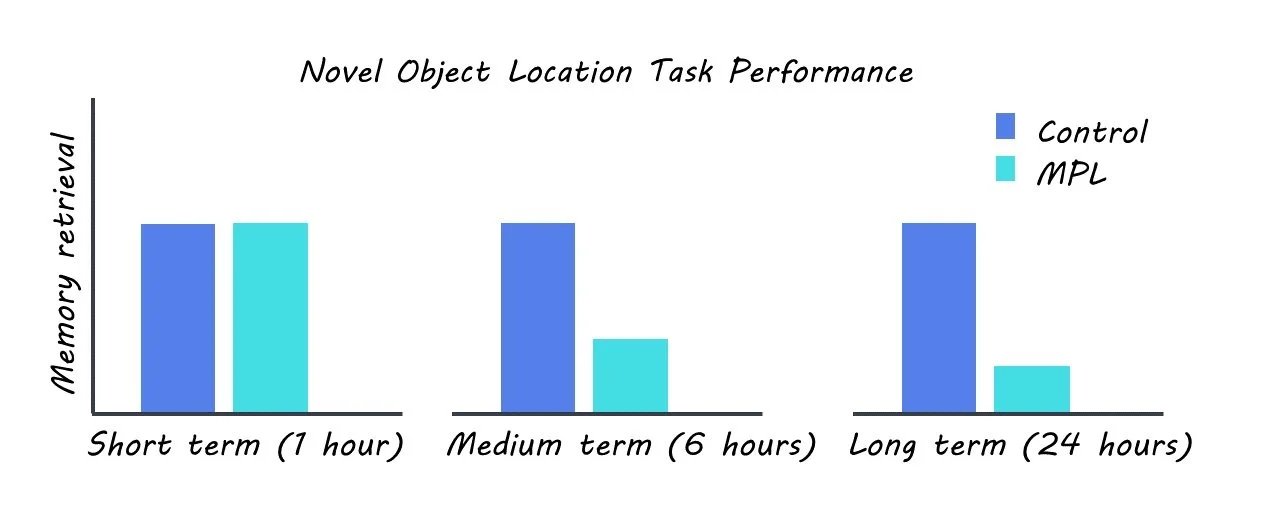
The authors used a rat model to mimic corticosteroid treatment by administration of the commonly prescribed corticosteroid methylprednisolone (MPL). The rats were housed in a 12-hour light-dark and were administered MPL orally. The authors screened for behavioral, genetic, and neurophysiological changes in the MPL group relative to a control group. They monitored changes in locomotion and body temperature between the two groups in order to screen for any potential impacts of the treatment on these behaviors. They also performed a novel object location task to assess changes in short, intermediate, and long-term memory. RNA sequencing was used to measure changes in gene expression in the two groups. Protein levels were assayed using Western blotting. Intracellular ex-vivo recordings were used to explore changes in synaptic plasticity as a result of corticosteroid treatment.

What did they find?

The authors observed that corticosterone treatment caused disruptions in synaptic physiology and gene expression in the hippocampus. Specifically, the authors found that corticosterone treatment impaired long-term potentiation (a measure of synaptic plasticity that is associated with long-term memory), altered the expression of genes involved in circadian regulation and synaptic plasticity, and disrupted the amplitude and frequency of miniature excitatory postsynaptic currents (mEPSCs) in the hippocampus. The authors also showed that corticosterone treatment inhibited NMDAR-dependent plasticity, which is a key mechanism underlying learning and memory in the hippocampus.

In addition, their behavioral task found that rats treated with corticosterone exhibited impaired intermediate and long-term memory, but not short-term memory. The authors further investigated the molecular basis for these effects by analyzing gene expression in the hippocampus. They found that corticosterone treatment dysregulated genes that are known to be crucial for memory processing in the hippocampus and circadian regulation, including CAMKII and CLOCK. Crucially, the authors found that the time of day was a potent modulator of hippocampal activity and that this temporal regulation is disrupted by corticosteroid treatment.

What's the impact?

This study sheds light on the underlying mechanisms of the cognitive side effects of long-term corticosteroid treatments, which are very commonly used to treat many inflammatory and immunologic disorders. Furthermore, these findings could lead to the development of new therapeutic approaches for patients who experience cognitive side effects of corticosteroid treatments. 

Access the original scientific publication here.

Using Virtual Reality to Probe Human Spatial Learning and Decision Making

Post by Anastasia Sares

The takeaway

Using a virtual reality environment and fMRI, researchers can study human navigation and how we can learn what is rewarding in our environment. This article shows how the orbitofrontal cortex, a region located in the frontal lobe just above the eyes, interacts with our hippocampus (the brain’s map-making region) in situations where knowledge about our surroundings can help when we face new decisions.

What's the science?

One of the difficulties in trying to understand how humans learn about objects in their environment is that objects close to each other in space also tend to be close to each other in time. That is, as we explore spaces, we tend to encounter close-by objects sooner and far-away objects later. However, since everyone navigates their environment in slightly different ways, they experience their own unique sequence of events while reconstructing the same space in their head.

This week in Nature Neuroscience, Garvert and colleagues designed a virtual reality experiment to pull apart the time and space aspects of our navigational abilities and to show what happens when one of those dimensions (here: space) can inform new decisions.

How did they do it?

The authors conducted a three-day experiment where participants learned their way around a virtual arena with monsters in it; the knowledge they acquired in the arena could later be used to maximize rewards.

On day 1, participants started by exploring the arena, which had some landmarks around the edge, and hidden monsters scattered about (they would not appear until the person “walked” to just the right position). The goal on the first day was just to memorize the locations of the monsters. After exploring, participants were tested by walking to the locations of the monsters they were tasked to find.

On day 2, participants had some additional practice and were put into the functional magnetic resonance imaging (fMRI) scanner. While their brains were scanned, they viewed images of the monsters and were encouraged to think about their location in the arena. Sometimes, two monsters would appear, and they would have to choose the one whose location was closest to the monster presented previously. This allowed the researchers to investigate how the monsters were represented in the brain.

On day 3, participants practiced locating the monsters again and entered the scanner for a second time. This time, they were presented with pairs of monsters and told to select the one with a higher reward, knowing that monsters that lived in close proximity would be worth a similar number of points. Points were presented after they made their choice on each trial. In reality, monsters close to a certain point in the arena were worth more points, and those further away were worth less. At the end of the choice task, participants were presented with two monsters whose reward value had never been shown in the choice task and asked to predict the reward value. Participants could successfully do this, demonstrating that they used knowledge about the monster locations to predict unseen rewards. Finally, participants completed the viewing task again, just like they had on day 2, this time thinking about both the spatial location and reward value of each monster.

What did they find?

Participants had a variety of exploration strategies in the virtual environment, and thus each person visited the monsters in a unique order.

However, in the choice task, the spatial relationships and not the order in which they saw the monsters could be used to maximize rewards. The authors compared different models to see which best explained the participants’ choices: one that relied only on predictions based on spatial relationships, one that relied on predictions from that person’s exploration order, and one that combined both. It was the “both” model that ended up making the best predictions. People use a combination of time (the order of their experiences) and space (the map representations they have built) to represent the relationship between two objects. Some people leaned more towards spatial relationships and were better at updating them, while others relied more on the order in which they had experienced things.

This was also true in the brain. The team focused on the hippocampus, a region that is known to help us make cognitive maps and navigate our environment. The authors were able to use activity in this region to predict how much people used their knowledge about the relative distances between monsters to infer the values of monsters whose values they had not experienced, yet, on day 3. So, this region is likely involved in helping people generalize about properties of their environment such as whether it’s rewarding or not. However, it was not the only brain region involved: activity in the orbitofrontal cortex was related to the participant’s ability to update and refine their strategy, focusing on spatial aspects to get the optimal reward. This is a neat confirmation of a similar animal-based result in a recent brainpost.

What's the impact?

Learning to navigate to rewards is a crucial skill for any animal’s survival—we must have maps to rewards like food sources or safe places to sleep. The interaction of the hippocampus and the orbitofrontal cortex seems to be key to this skill, now being shown in both animals and in humans.