The Link Between Phantom Ear-Ringing and the Brain’s Emotion Centers

Post by Anastasia Sares

The takeaway

Tinnitus is a condition of hearing sound in the absence of any external stimulus, usually a ringing, hissing, or roaring. Though it was initially thought to be purely a hearing disorder related to noise exposure and other physical factors, scientists have discovered surprising connections to emotional and psychological health.

Recently in the International Journal of Molecular Sciences, Singh and colleagues reviewed the literature linking tinnitus to the limbic system: a collection of structures located deep in the brain with functions like spatial memory, threat processing, motivation, and stress. These limbic system connections may explain why tinnitus is related to other aspects of mental health.

What is tinnitus?

There are two prominent theories about the causes of tinnitus. In the first one, lack of auditory input causes the fibers that normally carry auditory signals to fire spontaneously, generating waves of activity that have no outside source. In the second theory, reduced input leads to a lack of inhibition in key auditory areas, which causes an overall increase in auditory signals despite no external sound. In short, in the absence of stabilizing auditory input, brain regions processing sound start to behave differently, going so far as to “invent” signals and interpret them as sound.

Both of these theories highlight the fact that auditory information passes through multiple structures in the brain. Because so many brain regions are involved, there is also the potential for those brain regions to influence the experience of tinnitus. In fact, the experience of tinnitus encompasses both the phantom sounds as well as a person’s reaction to those sounds (conscious or subconscious).

How is the limbic system involved?

We’ll focus on a few prominent brain regions that have been studied in relation to tinnitus.

The hippocampus: this region is tightly tied to the auditory system since auditory information helps with spatial navigation, and the hippocampus is also responsible for long-term auditory memories. Brain scans of people with tinnitus show a decrease in the size of the hippocampus, and activity in the hippocampus can help predict the unpleasantness of people’s tinnitus experience. Since noise exposure can cause noticeable changes in the hippocampus, it may be a key intermediate region in the pathway from noise exposure to tinnitus.

The amygdala: the amygdala is involved in fear and other emotional processing; it also has a strong connection to regions that regulate incoming auditory information. Activity in the amygdala may be a key part of the process that causes stress and negative emotions to intensify the experience of tinnitus.

The nucleus accumbens and anterior cingulate cortex: These are structures involved in motivation, reward, and addiction, and they work against each other. The nucleus accumbens acts to arouse us and orient our attention to both pleasant and unpleasant stimuli, and more neural activity in this area seems to aggravate tinnitus. The anterior cingulate, on the other hand, can help us control and filter out unpleasant auditory stimuli, working against tinnitus symptoms. If this pathway is weakened, it would provide less filtering and thus, stronger symptoms.

The basal ganglia: The basal ganglia are small regions deep in the brain that form a hub for much of the brain’s activity. How the basal ganglia are involved in tinnitus is somewhat unclear, but there are some dramatic examples of how either stroke or deep brain stimulation in these regions can significantly reduce tinnitus.

What's the impact?

Effective treatments for tinnitus are few and far between, and there is no gold standard. Knowing that limbic regions are involved in creating or exacerbating these symptoms, we can make better guesses at what therapies might work, like drugs that target limbic areas primarily, or deep-brain stimulation in the basal ganglia. Hopefully, more limbic-system-focused therapies are on their way.

Access the original scientific publication here.

How The Hypothalamus Regulates Hormone Release During Fasting

Post by Baldomero B. Ramirez Cantu

The takeaway

Fasting activates agouti-related peptide (AgRP)-expressing neurons in the hypothalamus which disinhibit neurons in the ​​paraventricular hypothalamus (PVH). This process leads to the release of corticosterone, a hormone that helps manage glucose levels which provides energy during fasting.

What's the science?

During fasting, the body undergoes various essential survival responses, including the activation of the hypothalamic-pituitary-adrenal (HPA) axis, which increases the levels of stress hormones (e.g. cortisol in humans or corticosterone in rodents). These hormones prevent drops in blood sugar caused by fasting and maintain glucose balance. This response is crucial for preventing low blood sugar during fasting, and although its importance is recognized, the exact mechanism behind this activation has remained a mystery. This week in Nature, Douglass, Resch, Madara et al. delve into the underlying neural mechanisms and specific neuron-type roles in the activation of the HPA axis during fasting.

How did they do it?

The authors used a variety of techniques to investigate the role of the hypothalamus in regulating corticosterone release during fasting in mice. They primarily relied on plasma corticosterone measurements to measure HPA-axis activation, optogenetic and chemogenetic manipulations to probe the function of different cell types and to map connectivity between different hypothalamic regions, and ex-vivo preparations to assess the role of different receptor types in this pathway.

First, the authors confirmed previously reported data that fasting activates the HPA axis and increases corticosterone levels - by fasting mice for 24-hours and measuring their plasma corticosterone levels. The authors then used chemogenetics to activate or inhibit AgRP neurons and measured the effects on corticosterone levels. To further confirm the role of AgRP neurons in activating the HPA axis, the authors conducted experiments where they monitored the activity of a PVH-Crh (a specific subclass of PVH neurons crucial for initiating the release of corticosterone) by measuring their activity levels using fiber photometry, while simultaneously performing chemogenetic activation of AgRP neurons as a function of chemogenetic manipulation of AgRP neurons.

Next, they wanted to understand how AgRP neurons synaptically influence the activity of PVH-Crh neurons. Since AgRP neurons release inhibitory neurotransmitters and do not directly excite PVH-Crh neurons, they hypothesized that AgRP neurons might inhibit other neurons that in turn inhibit PVH-Crh neurons - thereby activating PVH-Crh neurons via reduced inhibition. They conducted experiments using ex-vivo electrophysiology, recording inhibitory currents onto PVH-Crh neurons while using receptor-specific agonists or antagonists (NPY and GABA). They also created genetic mutants of the NPY and GABA receptors in order to probe their role for PVH-Crh neuron inhibition in-vivo.

Finally, the authors wanted to identify the source of inhibitory GABAergic input that influences PVH-Crh neurons. They used a technique called retrograde rabies mapping to identify brain regions sending GABAergic signals to PVH-Crh neurons. Next, they employed an optogenetic-based method called channelrhodopsin assisted circuit mapping (CRACM) to confirm that neurons from a specific brain region inhibit PVH-Crh neurons ex-vivo, and fiber photometry to confirm that projections from this brain area to PVH are inhibited by AgRP neurons in-vivo.

What did they find?

The authors found that the activation of AgRP neurons increased corticosterone levels even in well-fed mice, while inhibiting these neurons suppressed the usual increase in corticosterone seen during fasting. This indicates that AgRP neurons play a crucial role in releasing corticosterone and are essential for this response during fasting. Chemogenetic activation of AgRP neurons drove rapid and sustained activation of PVH-Crh neurons while inhibition appeared to have the opposite effect. These results further support the role of AgRP neurons in this pathway, given the crucial role of PVH-Crh neurons in the release of corticosteroids and the activation of the HPA axis.

They also found that NPY and GABA can reduce inhibitory tone onto PVH-Crh neurons through receptors located on GABAergic afferents in their ex-vivo preparation. Through in-vivo experiments in genetically modified mice, they discovered that both NPY and GABA are not individually necessary for AgRP neurons to activate the HPA axis, but their combined effect is crucial. The study suggests that GABA release from AgRP neurons acting on GABA-B receptors on GABA-ergic afferents to Crh neurons in the PVH is necessary for activating the HPA axis.

Finally, the authors identify the bed nucleus of the stria terminalis (BNST) as the source of tonic inhibition to PVH-Crh neurons. Chemogenetic inhibition of inhibitory BNST neurons increased plasma corticosterone levels, indicating that inhibiting these neurons stimulates the HPA axis. Specifically inhibiting the BNST → PVH pathway also stimulated the HPA axis. Additionally, their fiber photometry results showed that stimulation of AgRP neurons suppressed the synaptic activity of BNST axon terminals in the PVH. Overall, their findings suggest that inhibitory afferents from the BNST normally suppress PVH-Crh neuron activity and that during fasting, AgRP neurons inhibit these afferents, reducing GABAergic tone onto PVH-Crh neurons and stimulating the HPA axis. 

What's the impact?

Understanding how neurons in the hypothalamus influence the body's adaptive responses to energy deficit and stress is paramount to providing insights into potential therapeutic targets for managing conditions related to metabolic and hormonal imbalances. These results help us gain a better understanding of the neural mechanisms by which AgRP neurons play a pivotal role in activating the HPA axis during fasting.

The Impact of Deep Brain Stimulation on Brain Networks and Treatment Outcomes in Treatment-Resistant Depression

Post by Kulpreet Cheema

The takeaway

Deep brain stimulation of the subcallosal cingulate gyrus in patients with depression induces changes in brain network properties, which is correlated with improvements in depression symptoms.

What's the science?

Treatment-resistant depression (TRD) is a severe form of depression that does not respond to standard treatments. Deep brain stimulation (DBS) is an emerging therapy that involves implanting electrodes in specific brain regions to modulate neural activity and potentially alleviate depressive symptoms. The effectiveness of DBS varies among patients, and personal factors might be the key to understanding this variability. This week in Molecular Psychiatry, Ghaderi and colleagues aimed to understand how DBS affects brain networks in TRD patients using electroencephalography (EEG), a non-invasive technique that measures the brain’s electrical activity.

How did they do it?

The study involved twelve TRD patients who underwent subcallosal cingulate gyrus (SCG) DBS. The researchers collected resting-state EEG data and Hamilton Depression rating scale data from participants at three sessions: before surgery, one to three months after surgery, and six months after surgery. During the second and third sessions, EEG data was collected with DBS turned on and off sequentially.

To analyze brain network properties, the researchers used an analytical method known as graph theoretical analysis to decipher whether there were distinct differences in brain network properties between patients who responded positively to the DBS treatment (responders) and those who did not (non-responders). Using EEG to study oscillatory activity in brain networks, the authors analyzed four different frequency bands — the delta and alpha bands were of particular interest due to their relevance to depression.

What did they find?

The study revealed several important findings. First, the baseline brain network properties in patients — especially in the delta and alpha frequency bands — were associated with the outcomes of DBS treatment. Responders to DBS showed lower levels of network segregation, integration, and synchronization at baseline compared to non-responders. Second, DBS led to changes in brain network properties over time, characterized by increased integration and synchronization of brain regions. These changes were particularly evident in the delta frequency band.

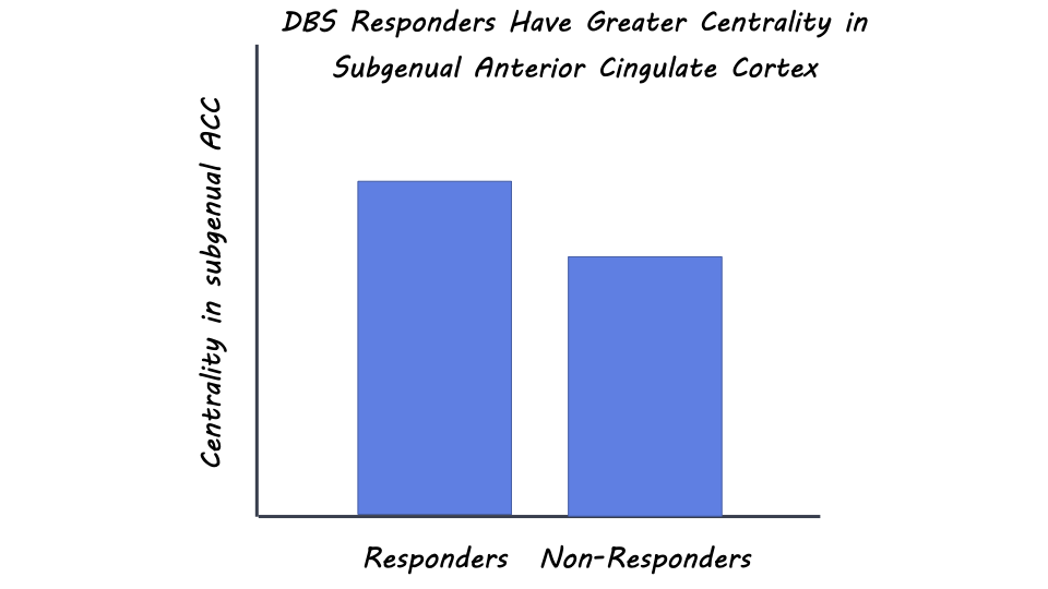
Third, the researchers found that responders had higher centrality - a measure of the importance of a node in a network for information propagation - in the subgenual anterior cingulate cortex (ACC), a brain region associated with depression symptoms. DBS led to a reduction in centrality in this region, which correlated with treatment response. Other brain regions, like the primary somatosensory cortex and parahippocampal gyrus, also showed alterations in centrality, indicating their involvement in TRD and DBS response.

What's the impact?

This study sheds light on the complex effects of DBS on brain networks in treatment-resistant depression. By using EEG and advanced analytical techniques, researchers identified specific brain network features associated with treatment response and demonstrated how DBS induces changes in these networks over time. The findings provide valuable insights into the potential mechanisms underlying the therapeutic effects of DBS in TRD patients. This knowledge could contribute to the development of personalized treatments for individuals with treatment-resistant depression, ultimately offering new hope for improved mental health outcomes. 

Access the original scientific publication here