How Engrams Become Selective During Memory Consolidation

Post by Shireen Parimoo

The takeaway 

Engrams are ensembles of neurons that are activated during memory encoding and retrieval. During memory consolidation, excitatory hippocampal neurons facilitate the reorganization of engrams while inhibitory interneurons regulate the selectivity of those engrams for specific memories.

What's the science?

Engrams are ensembles of neurons in the hippocampus that are activated during learning and memory encoding. When those memories are later retrieved, at least a portion of the same neurons are reactivated, but it is unclear whether the engram from the learning period remains intact throughout consolidation (i.e., static engram) or if new neurons are incorporated into the engram while some of the original neurons are removed (i.e., dynamic engram). This week in Nature Neuroscience, Tomé and colleagues used computational modeling and contextual fear conditioning experiments in mice to investigate the neurobiological mechanisms by which engrams evolve as fear memories are consolidated in the hippocampus.

How did they do it?

The authors created a neural network model of the hippocampus that included inhibitory and excitatory neurons. They simulated memory encoding by providing a training stimulus as input to the network from a separate population of cells. Hippocampal neurons with a spiking rate above a specified threshold were considered to be part of the engram ensemble. In the consolidation and probing phases, the training stimulus was presented randomly to facilitate memory selectivity and to examine memory reactivation, respectively. Lastly, the training stimulus and novel stimuli were presented to the hippocampal network during recall. The recall rate (firing rate of engram cells in response to the training vs novel stimuli) and engram cell overlap during training and later memory reactivation were used to track the evolution of the engram ensemble. The activity of inhibitory and excitatory neurons was also manipulated to generate predictions about their role in engram evolution and memory retrieval.

Mice underwent contextual fear conditioning and their freezing behavior was recorded in the training environment during the learning phase, in a neutral environment, and again in the training environment during recall. Optogenetic and chemogenetic techniques were used to inhibit hippocampal neurons, along with activity-dependent cell labeling and calcium imaging to identify (i) engram composition during learning, and (ii) the role of inhibitory and excitatory neurons in memory consolidation and retrieval. Comparing the training context during recall to the neutral context, the amount of freezing behavior was used to determine memory selectivity while the overlap in activated engram cells was used to track engram evolution.

What did they find?

In the network model, the overlap between training-activated neurons and those activated during the probing phase decreased, indicating that the composition of the engram ensemble changes over time. The engrams also became more selective over time as their recall rate only dropped below the threshold for the novel stimuli. Altogether, the network model predicted that engrams change dynamically and become more selective over the course of memory consolidation. Importantly, the potentiation of inhibitory synapses between the inhibitory engram neurons and both engram and non-engram cells drove the dynamic reorganization of the engram during consolidation. That is, the activity of inhibitory neurons controlled which hippocampal neurons would be added to or removed from the initial engram ensemble.

Blocking long-term potentiation of excitatory neurons during consolidation led to a stabilized or static engram and impaired recall in the network model. On the other hand, blocking inhibitory interneurons in the neural network during consolidation and recall disrupted memory selectivity but did not affect engram reorganization. In mice, inhibiting the inhibitory neurons after fear conditioning and during recall similarly impaired memory selectivity as the mice could not discriminate between the training and neutral contexts. Together, these results show that during the memory consolidation phase, excitatory hippocampal activity is important for the dynamic reorganization of engrams while inhibitory activity underlies memory selectivity.

What's the impact?

This study revealed that engrams are dynamic and undergo reorganization during memory consolidation, which in turns facilitates memory selectivity for successful recall. Future work can build on these findings to provide a better understanding of the neurobiological basis of memory-related pathologies.

Access the original scientific publication here.

Exercise and Movement-Triggered Anxiety Relief

Post by Baldomero B. Ramirez Cantu

The takeaway

This study elucidates the role of a previously unidentified neural circuit involving the cerebellum, amygdala, and hypothalamus in driving the anxiolytic effects of exercise. 

What's the science?

Anxiety disorders constitute a spectrum of mental health conditions marked by enduring sensations of fear, apprehension, or discomfort. Exercise is hailed as a first-line and often successful intervention for managing anxiety symptoms. Despite the well-documented relationship between physical activity and anxiety reduction, the exact neural mechanisms underlying exercise's impact on anxiety remain incompletely understood.

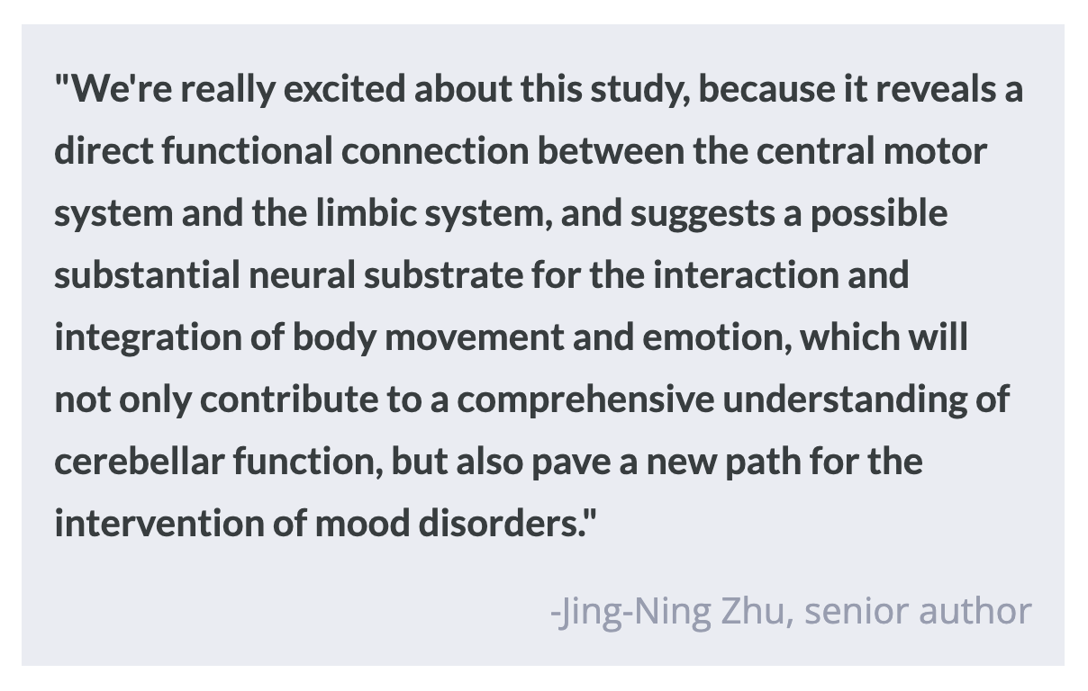
The amygdala and hypothalamus are known to be key nodes in the brain's "anxiety circuitry”, contributing to the manifestation and regulation of anxiety-like behaviors. However, a much less explored brain region in this context is the cerebellum, a region primarily associated with motor function. Advancing our understanding of the interaction between anxiety and motor-associated brain regions could offer significant insights into how exercise modulates anxiety. This week, Zhang, Wu, Shen, Ji, et al. published a study in Neuron that investigates the functional connectivity among the cerebellum, amygdala, and hypothalamus in the context of exercise-induced anxiety relief, and provides one of the first pieces of direct evidence of cerebellar involvement in anxiety modulation. 

How did they do it?

First, functional magnetic resonance imaging (fMRI) data were collected from humans to assess the functional connectivity between the cerebellum and amygdala as it relates to anxiety. This human study served as a starting point, establishing a relationship between the cerebellar-amygdala circuit and anxiety. Next, to study this circuitry with greater precision and understand the specific subregions and cell types involved in the exercise-induced amelioration of anxiety, the authors performed experiments in rats as model organisms.

Viral mediated tracing techniques, including anterograde and retrograde tracer injections, were used to map the direct projections from the dentate nucleus of the cerebellum to the central lateral amygdala. These experiments aimed to delineate the anatomical connectivity between cerebellar nuclei and amygdalar subregions, shedding light on the structural basis of cerebello-amygdalar circuitry involved in anxiety regulation. Viral tracing experiments were also performed in order to map the connectivity between the DN and orexin-expressing neurons in the perifornical area of the hypothalamus. The authors conducted fMRI scans on anesthetized rats to investigate the cerebellar-amygdala circuitry's functional connectivity between rats subjected to exercise and controls. 

Finally, optogenetic and chemogenetic manipulations were employed in rats subjected to anxiety assays following rotarod running to elucidate the impact of motor activity on anxiety levels. These manipulations allowed researchers to selectively activate or inhibit cerebellar nuclei neurons projecting to the amygdala, providing insights into the causal relationship between cerebello-amygdalar circuitry and anxiety symptoms.

What did they find?

First, the researchers observed anxiety was negatively correlated with cerebello-amygdalar functional connectivity in humans. Next, they found reduced anxiety levels in rats subjected to rotarod running. For example, an increased preference for open arms in maze tests was observed (indicating an anxiety reduction), demonstrating that exercise had similar effects on the anxiety responses of rats and humans. Notably, functional connectivity analyses of rat fMRI scans revealed enhanced connectivity between the cerebellar nuclei and amygdala following motor activity.

They identified a direct cerebello-amygdalar projection, particularly from the dentate nucleus to the central lateral amygdala, suggesting a structural basis for regulation of anxiety-like emotions. Optogenetic and chemogenetic manipulations demonstrated that activation of cerebello-amygdalar projections significantly reduced anxiety-like behaviors in rats, while inhibition of this pathway attenuated the anxiolytic effects of exercise. Additionally, the researchers uncovered orexinergic hypothalamo-cerebellar projections as crucial components in anxiety regulation, particularly during challenging exercises. Activation of orexinergic neurons in the perifornical area of the hypothalamus alleviated stress-induced anxiety, highlighting the role of hypothalamo-cerebellar circuits in modulating anxiety behavior.

What's the impact?

This study significantly advances our understanding of the neural circuitry underlying anxiety regulation, particularly highlighting the previously unknown intricate interplay between the cerebellum, amygdala, and hypothalamus. This information is invaluable to our advancement of treatments and interventions for anxiety disorders.

Biomarkers for Diagnosing Alzheimer’s Disease in the Indigenous Population

Post by Soumilee Chaudhuri

The takeaway

This study reveals unique patterns of blood-based clues linked to Alzheimer’s Disease (AD) in older American Indian (AI) individuals — a critically understudied population in healthcare and biomedical science. Blood-based biomarkers in this population suggest early and more widespread presence of AD-related changes in this indigenous cohort, compared to other ethnic groups.

What's the science?

Alzheimer’s Disease (AD) is the most common form of dementia and affects over ~6.5 million people in the United States. As a major public health concern, early identification of AD is important for effective prevention and treatment strategies for this debilitating disorder. However, existing methods of diagnosis such as cerebrospinal fluid (CSF) analysis and positron emission tomography (PET) imaging are invasive, expensive, and unaffordable for minoritized populations, including American Indians for whom treatment is often inaccessible due to socioeconomic and systemic factors. Therefore, there is a critical need for noninvasive and low-cost biomarkers that can accurately detect AD pathology, particularly in these underserved communities. The Strong Heart Study (SHS) addresses this gap by investigating blood biomarkers related to AD in a cohort of older American Indian individuals recruited from designated field centers in Arizona, North & South Dakota, and Oklahoma. By examining associations between these biomarkers and clinical, imaging, and cognitive findings, the authors provide critical insights into AD characteristics, diagnostics, and risk factors specific to the American Indian elderly population.

How did they do it?

Initially, SHS recruited American Indian adults aged 64-94 years old from tribal lands in the US Northern Plains, Southern Plains, and Southwest starting in 1981-1991; thereafter, the survivors of this initial cohort were recruited for cognitive aging and AD studies in 2010 ( N = 818) and invited back for a second visit in 2017 (N = 403). Five blood biomarkers of AD including phosphorylated tau (ptau), amyloid beta (A), glial fibrillary acidic protein (GFAP), and neurofilament light chain (NfL) were obtained from all participants using designated research platforms. Participants also underwent magnetic resonance imaging (MRI) and neuropsychological & cognitive testing (such as memory, executive function, simple and divided attention, etc.). Researchers used statistical methods such as regression analyses and Receiver Operator Characteristics (ROC) to assess the relationships between these blood biomarkers and various imaging measures and cognitive outcomes.

What did they find?

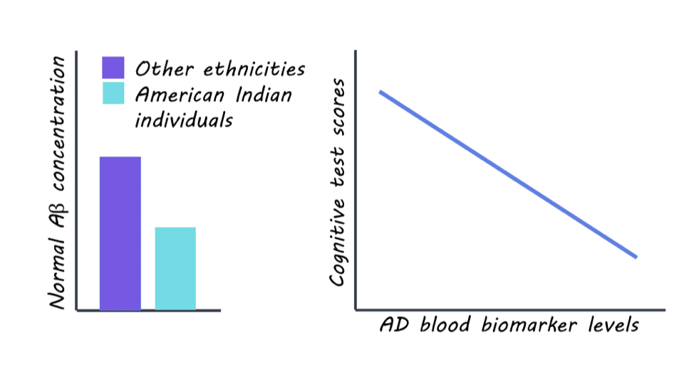
The researchers found significant differences in blood biomarker levels related to AD pathology among the SHS cohort of older American Indian individuals compared to other ethnic groups. The levels of normal Amyloid beta (A) — the lack of which is a hallmark of AD pathology — were significantly lower in this cohort compared to non-Hispanic white (NHW), African American (AA), and Hispanic/Latino populations, indicating a greater degree (almost 3 times) of pathology in this cohort compared to other populations. However, the levels of GFAP and NfL — other biomarkers of AD — were similar in this American Indian cohort as compared to other age-comparable populations. Associations were observed between blood biomarker levels of AD and MRI and cognitive test scores, suggesting potential implications for AD diagnosis and risk assessment in this population. Taken together, these findings reflect that previous studies of comparable risk between American Indian and non-Hispanic White individuals may underestimate AD prevalence in American Indians.

What's the impact?

This is the first study to investigate blood biomarkers for Alzheimer’s Disease in a population historically underrepresented in research: American Indians or the Indigenous people. These findings highlight the importance of understanding cognition and aging in diverse populations facing healthcare disparities so as to improve diagnosis and craft efficient, non-invasive, and affordable treatment strategies. It also shows us that previous studies have severely underestimated the risk of AD in this vulnerable population and urges us to be mindful of recruiting diverse participants from all demographics in AD studies so as to tailor diagnosis and treatment strategies to all.  

Access the original scientific publication here