Tau Protein in Human Neurons is Essential for Amyloid Beta-Driven Toxicity in Alzheimer’s Disease

Post by Laura Maile

The takeaway

Amyloid beta and tau are two proteins that aggregate in the brain of patients with Alzheimer’s Disease and are linked to the degeneration of neurons and symptoms of the disease. When tau is depleted in human neurons, hyperactivity and neurodegeneration caused by amyloid beta is reduced.  

What's the science?

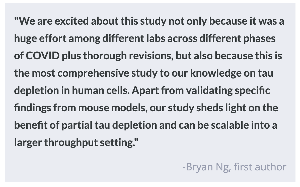
Alzheimer’s Disease (AD) is characterized by the buildup of plaques and tangles, which are aggregates of amyloid beta and tau proteins, among other pathologies. One of the prominent theories of the primary cause of AD, the “amyloid hypothesis,” states that the buildup of amyloid beta leads to toxicity of neurons and neurodegeneration.  Though the physical pathology of the disease has been well-described, scientists do not yet fully understand the interplay between amyloid beta, tau, and neurodegeneration, especially in human experimental models. Mouse models with tau genetically knocked out have shown that amyloid beta toxicity is dependent on tau. This week in Molecular Psychiatry, Ng and colleagues used gene-edited human cells to deplete tau and examine its role in amyloid beta-driven toxicity. 

How did they do it?

The authors used Crispr-Cas9 to genetically modify the gene for tau in human induced pluripotent stem cells (hIPSCs). hIPSCs are generated using human cells that are induced back into being stem cells and then differentiated into specific cell types, such as neurons. Using this method, the authors could disrupt the tau gene in human cells. They confirmed that they had effectively depleted the tau transcript and protein in the cortical neurons generated from hIPSCs. They first examined the effects of tau depletion on neuronal activity, using electrodes to measure extracellular field potentials. Using the same methods, they measured neural activity over time in cells treated with homogenate from post-mortem AD brain tissue. To determine the specific effects of amyloid beta on synapse loss seen in AD, they extracted amyloid beta from post-mortem brains of AD patients and treated neurons both with and without tau. They then examined the effects on synapse loss using immunocytochemistry to label synaptic markers. Next, the authors investigated the effect of tau loss on the movement of mitochondria down axons by plating the hIPSC-derived neurons on one side of a chamber and live imaging the movement of mitochondria. This was followed by a similar experiment where they added amyloid beta oligomers to the neurons to mimic a toxic amyloid beta insult, and monitored mitochondrial movement. Finally, they tested whether the depletion of tau could protect against neurodegeneration caused by amyloid beta. To do this, they again used amyloid beta oligomers to provide a toxic insult to hIPSC-derived cortical neurons with and without tau, and compared cell death in each set of neurons.

What did they find?

The authors discovered that cortical neurons with the tau protein depleted showed reduced neuronal activity. Neurons with tau that were treated with homogenate from the brains of AD patients showed hyperactivity over time, but both neurons without tau and neurons treated with an amyloid beta blocker were protected from this hyperactivity. This means that the hyperactivity observed was caused by amyloid beta and was dependent on tau. Treatment with amyloid beta from AD brains leads to synapse loss in normal neurons, but not in tau-deficient neurons, meaning that tau is essential for amyloid beta-induced synapse loss. When normal neurons were treated with amyloid beta, axonal transport of mitochondria was reduced. This effect was reversed in neurons missing tau, while the movement of mitochondria was not changed in cells missing tau that had not been treated with amyloid beta. This suggests that the dysfunction in mitochondrial transport caused by amyloid beta is dependent on tau. When regular hIPSC-derived cortical neurons were treated with amyloid beta, cell death increased. Partial and full depletion of tau reduced this amyloid beta-driven neurodegeneration, suggesting that even partial reduction of tau can prevent cell death. 

What's the impact?

This study found that amyloid beta-driven neuronal hyperactivity, synapse loss, axonal transport dysfunction, and neurodegeneration are dependent on tau in human cells. This suggests the continued importance of generating treatments to decrease tau in patients with AD.

How the Brain Accumulates Evidence to Make a Decision

Post by Natalia Ladyka-Wojcik

The takeaway

Intracranial electroencephalography was used in a large group of pre-surgical epilepsy patients to identify where in the brain perceptual decision-making begins and to show how brain activity might accumulate to support the strength and accuracy of decisions.

What's the science?

Perceptual decision-making involves selecting one choice from a set of alternatives based on incoming information from our senses. For example, a football player is engaged in perceptual decision-making when judging which player to pass the ball to during a game. When we make these decisions, our brain is also planning the corresponding motor actions, like the player gripping the football behind his head to throw it to his teammate. Recent research has identified parts of the brain involved in making these decisions, even in complex situations when the appropriate motor action might not be known in advance, but it remains unclear where exactly in the brain this process actually starts. This week in Nature Human Behavior, Gherman and colleagues pinpoint the signals in the brain responsible for these abstract decision-making processes.

How did they do it?

Previous research has found that brain activity linked to decision-making builds gradually over time, proportionally to the strength of incoming sensory information. As this evidence accumulates, it reaches a fixed threshold just before the animal makes a response and this threshold predicts both the animal’s choice and response time. In human neuroimaging research where resolution at the single neuron level is often not feasible, past studies have instead relied largely on functional magnetic resonance imaging (fMRI) to investigate brain regions involved in perceptual decision-making. However, fMRI has low temporal resolution, meaning that it doesn't show changes in brain activity quickly enough to demonstrate if there is a signal related to evidence accumulation. To overcome this limitation, the authors measured brain activity directly using intracranial EEG in a group of pre-surgical epilepsy patients who were asked to perform different perceptual decision-making tasks. This technique allows for measurement of a larger portion of the brain compared to single-neuron recordings and with better temporal resolution than fMRI.

First, the authors showed patients two simultaneous random-dot patches and asked patients to report the direction of the moving dots. The researchers measured the patients’ responses and the time it took to press a button to make their decision while recording their brain activity using intracranial EEG. Some patients also had to do the same task but with verbal responses after a delay, helping the researchers find brain activity related to perceptual decision-making, not just motor responses.

What did they find?

The authors found a widely distributed network of brain regions associated with perceptual decision-making, including the prefrontal cortex, parietal cortex, as well as inferior temporal and insular regions. Importantly, this network of brain regions showed high-frequency activity consistent with evidence accumulation in decision-making (whereas lower-frequency activity tends to be associated with motor preparation signals). This work suggests that activity in these regions gradually builds up until enough evidence is accumulated to make a decision (here, in response to the direction of dots) even before the patient presses a button or verbalizes their decision. The authors observed a gradual buildup of activity following the onset of sensory evidence at a rate that scaled with the strength of that sensory evidence. This activity reflected both the choice accuracy and response times of patients.

What's the impact?

This study found that activity in a distributed network of brain regions accumulates in response to sensory information to enable perceptual decision-making. It is the first to use intracranial EEG to directly measure high-frequency activity in these regions in humans, enabling an investigation into brain responses for decision-making even before motor response planning

Access the original scientific publication here.

Neuroscience-Backed Strategies to Help You Learn More Effectively

Post by Rachel Sharp

What is learning?

Whether we are in school, with our friends, or pursuing hobbies, we are always learning. But what exactly is learning? And why is it that we can pick up our favorite musician's new song after just a few listens, yet struggle to recall the contents of the Bill of Rights? Biologically, learning is the process by which neurons form and strengthen connections between each other. Every thought and decision we make requires communication between neurons across the brain. Picture groups of neurons working together, firing in unison to produce a thought, idea, or reaction. As these neurons fire together, their connections strengthen, making it more likely they'll fire together in the future. Forming new neural connections, strengthening existing ones, and pruning away unnecessary ones are the building blocks of learning, and understanding how to engage these processes can help us become more effective learners.

Learning occurs in three stages: encoding, storage, and retrieval. Let's explore what happens during each stage, both biologically and practically, and identify strategies to make these processes faster, more accurate, and longer lasting.

Effective encoding, storage and retrieval

Encoding refers to the initial process of encountering and interpreting new information. Our brains are constantly bombarded with information, so when it comes to learning, encoding is highly selective. Most of this filtering happens automatically based on the perceived importance of the information. Here are some strategies to enhance encoding:

1)    Direct Attention: Directing attention to relevant information and minimizing distractions helps strengthen encoding. This might involve highlighting key points or using different colors for important information.

2)    Attach Meaning: Attaching meaning to new information, especially by connecting it to what we already know, helps with encoding. Providing examples or relating topics to personal experiences makes it easier for the brain to process and retain information.

3)     Multiple Modalities: Absorbing information visually, verbally, and actionably recruits multiple different parts of the brain during the encoding process. Effectively, this creates multiple pathways by which the information can be retrieved later.

Storage involves the processing and maintenance of encoded information in the hippocampus. As you learn, neural connections will be formed, rearranged, and lost. So how information is stored, and how strongly it’s stored and maintained, will impact whether learned information persists over time. Varied repetition is key to improving storage, as it strengthens neural connections associated with the information. When you encounter information again and again, the associated neural connections will activate and become stronger. By diversifying repetition through different modalities, contexts, or examples, we ensure robust storage and easier retrieval.

Retrieval is the process of accessing and recalling stored information. It strengthens existing connections and is crucial for long-term memory retention. Now, let's explore some neuroscience-backed learning strategies that enhance encoding, storage, and retrieval.

What are some examples of neuroscience-backed learning strategies?

1.     Orchestrated Immersion: This involves fully immersing learners in their own learning experience. Activities like brainstorming sessions, relating new information to personal experiences, and engaging with thought-provoking questions enhance focused attention and the perceived value of the information, thus improving encoding.

2.     Relaxed Alertness: Creating a fun yet challenging learning environment can aid memory retention. Teaching through songs, dances, or competitive activities triggers positive emotions, diversifies neural connections, and promotes recall.

3.  Active Processing: Encouraging independent learning through group discussions and seeking out additional information strengthens knowledge retention. Engaging in contextual discussions with diverse perspectives deepens understanding and enhances memory consolidation.

Understanding how our brains learn can significantly improve our learning outcomes. By employing effective encoding, storage, and retrieval strategies rooted in neuroscience, we can improve our ability to acquire and retain information. From directed attention, meaningful engagement, and varied repetition, to immersive learning experiences and active processing, incorporating these strategies into our learning routines can make the journey of acquiring knowledge more efficient and enjoyable.

References +

Abdullah, Z., Istiqomah, T., & Sari, R. (2022). NEUROSCIENCE-BASED BIOLOGY SCIENCE LEARNING STRATEGIES AT THE ELEMENTARY SCHOOL LEVEL. Proceeding International Conference on Islam and Education (ICONIE), 2(1), Article 1.

BrainWare. (2020, October 10). Neuroscience of Learning Brain-Based Learning Strategies. Cognitive Literacy Solutions. https://mybrainware.com/blog/brain-based-learning-strategies/

Colvin, R. (2016). Optimising, generalising and integrating educational practice using neuroscience. Npj Science of Learning, 1(1), Article 1. https://doi.org/10.1038/npjscilearn.2016.12

Jamaludin, A., Henik, A., & Hale, J. B. (2019). Educational neuroscience: Bridging theory and practice. Learning: Research and Practice, 5(2), 93–98. https://doi.org/10.1080/23735082.2019.1685027

Memory (Encoding, Storage, Retrieval). (n.d.). Noba. Retrieved February 25, 2024, from https://nobaproject.com/modules/memory-encoding-storage-retrieval