Identifying Aging Subtypes with Machine Learning

Post by Lani Cupo

The takeaway

Three patterns of brain aging were identified in a large dataset of cognitively normal adults by a semi-supervised machine-learning algorithm: one subtype associated with typical aging, one with vascular factors, and one with higher levels of brain atrophy. The patterns were associated with different genetic backgrounds, lifestyle risk factors, and later cognitive decline. 

What's the science?

The process of aging is associated with many changes in neuroanatomy, some of which emerge before noticeable pathology, such as cognitive impairment or memory loss. Variables such as genetic background, demographics, and lifestyle factors can impact these neuroanatomical changes. Identifying subtypes of age-related changes early on could help clinicians to identify and intervene in pathological aging processes, however, early changes may be subtle and require large datasets to accurately identify. This week in JAMA Psychiatry, Skampardoni and colleagues investigated whether there are aging subtypes.

How did they do it?

By pooling data from 11 neuroimaging studies, the authors assembled 58,113 scans from 27,402 individuals aged 45-85. They used several metrics to assess brain health including regional volume and white-matter hyperintensity (WMH) volume (a measure of changes in the brain vasculature). First, the authors used principal component analysis (a data reduction technique) to identify participants with the lowest atrophy levels at each age group (45-55, 55-65, 65-75, and 75-85). This “resilient aging” group (A0) was used as a reference to identify subtypes of aging. Then, the authors used a machine learning algorithm known as Semi-Supervised Clustering via Generative Adversarial Networks (Smile-GAN) to identify subtypes with different trajectories of aging relative to A0. The subgroups were first identified in each age group but then assessed for consistency in trajectories across age groups. Then, the subgroups were compared for differences in genetics, clinical, and cognitive features. A subset of the full dataset included longitudinal data, allowing the authors to examine whether the identified groups were associated with long-term outcomes.

What did they find?

The authors identified three subtypes of aging consistent across the age groups. The first group, A1, showed mild atrophy mostly localized near the Sylvian fissure (the noticeable sulcus, or crease, that can be seen when viewing a brain from the side). The second group, A2, was associated with hypertension, showed greater atrophy, and had the highest burden of WMH. The third group, A3, showed more severe and dispersed atrophy across the brain and was associated with greater cognitive decline. Because group A1 was the largest with the least severe atrophy, it was considered typical aging. Older individuals in both groups A2 and A3 showed worse cognitive scores than groups A0 and A1, with A2 showing more cardiovascular risk factors and genetic risk factors for Alzheimer’s Disease, and group A3 showing the greatest levels of depression. Of note, these cognitive differences were still considered “sub-threshold”, as the included participants had no diagnosis. Groups A2 and A3 showed the greatest longitudinal cognitive decline, with group A2 showing the greatest progression of WMH.

What's the impact?

This study identified generalizable subtypes of aging identifiable from mid-life. By highlighting two trajectories of atypical aging and characterizing them in terms of cognitive, lifestyle factors, and genetic phenotypes, as well as long-term outcomes, this study shows groups that may be differentially susceptible to neurodegenerative disorders.

 Access the original scientific publication here.

The Complementary Roles of Dopamine and Serotonin in Decision-Making

Post by Shireen Parimoo

The takeaway

People are more likely to reject unfair monetary offers when they come from humans compared to computers. In the brain, serotonin levels are sensitive to the value of the offer itself while dopamine levels are sensitive to the change in the offer value from the previous trial as well as to the social agent providing the offer. 

What's the science?

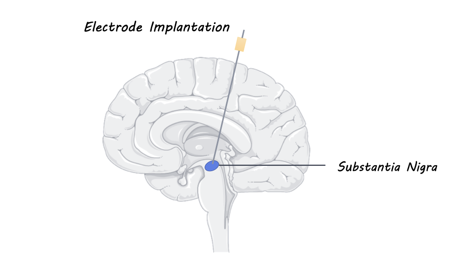
Neuromodulators play an important role in guiding behavior and decision-making. Dopamine, for example, is known to be important for reward-based processing. Pharmacological studies have provided insight into the causal role of neuromodulators like dopamine and serotonin in various social contexts. However, it is difficult to study the mechanisms through which they act because non-invasive brain imaging techniques like fMRI are limited in their spatial and temporal resolution. When given a drug that increases serotonin levels in the brain, for example, it is difficult to pinpoint precisely where and how long it takes for it to act in the brain. This week in Nature Human Behavior, Batten and colleagues used deep brain electrode recordings to monitor neuromodulator fluctuations in a region of the brainstem called the substantia nigra to understand the roles of dopamine and serotonin during social decision-making.

How did they do it?

The authors recruited four patients with Parkinson’s disease who played the ultimatum game while undergoing brain surgery across two sessions. For clinical purposes, the patients had electrodes implanted into the substantia nigra pars reticulata, which receives both dopaminergic and serotonergic inputs from other regions of the brain. Using a statistical model (convolutional neural network) applied to the electrochemical currents recorded from the electrodes, and knowledge of how dopamine and serotonin concentrations typically change this signal, the researchers were able to estimate changes in these neuromodulators with high temporal precision.

The ultimatum game is a social fairness game in which participants are offered a certain split of a monetary amount (e.g., 40% of $20). If they choose to accept the offer, then the money is split accordingly (i.e., the participant receives $8) but if they reject the offer, then no one receives any money. Participants played against the computer player that either had a human or a computer avatar and made offers valued between $1-9 that participants could accept or reject. The authors examined whether participants were more or less likely to accept the offers based on the value of the offer, human vs. computer avatar condition, and offer history, which is the change in the value of the offer from the previous trial. They also investigated how dopamine and serotonin levels fluctuated after they were given an offer. Specifically, neuromodulator levels were examined with respect to participants’ decisions, the offer values, offer history, and across the two avatar conditions. 

What did they find?

Participants were more likely to accept larger offers (i.e., $9 vs $3 offer) and offers made by the computer avatar but offer history did not affect their decision to accept or reject the offer. Participants also took longer to reject than to accept offers overall. Neither dopamine nor serotonin levels were altered by the decision to accept or reject an offer, but dopamine increased when the offer came from the human avatar compared to the computer avatar. This suggests that dopamine tracks social context (i.e., avatar condition) but not necessarily the decision itself. Dopamine was also sensitive to offer history, as it decreased when the value of the offer dropped but increased when the offer value increased. Serotonin, on the other hand, was sensitive to the value of the offer itself, but not to offer history. That is, serotonin levels were higher when the offers were high and lower in response to lower offers. Altogether, these findings show that serotonin is involved in value-based processing while dopamine is important for social context and for tracking the change in offer value across trials. 

What's the impact?

This study is the first to use electrochemistry in the awake human brain to demonstrate the differential roles of dopamine and serotonin in value-based processing in a social context. This study not only advances our understanding of neuromodulators in decision-making processes but also showcases the utility of electrochemistry as an exciting new method for human neuroscience research. 

Access the original scientific publication here.

How Listening to Live and Pre-Recorded Music Changes Brain Activity

Post by Meredith McCarty

The takeaway

Listening to music is not only an enjoyable and common pastime but has been found to correlate with changes in brain activity in many key regions involved in emotional processing. Live music is found to be more closely correlated with increased activity in key brain networks involved in emotional processing than pre-recorded music.

What's the science?

Music listening evokes strong feelings in listeners and has also been found to correlate with increased activity across the affective brain network which is involved in emotional processing and emotional recognition. Key regions associated with this affective network are the limbic system, most notably including a brain region called the amygdala, which is involved in emotional processing and processing of music-evoked emotions. Because of the influence of music listening on activity in this affective brain network, music can be a powerful tool to help us better understand brain dynamics. This week in PNAS, Trost and colleagues use a novel closed-loop music performance experimental setup in order to better understand the dynamics of music listening in the brain.

How did they do it?

To study how music listening affects brain activity, the researchers designed a novel closed-loop music performance setup, where the musician and the listener are influencing each other in real time. The researchers recruited 27 participants who had no musical experience to listen to different music through in-ear headphones while in a functional magnetic resonance imaging (fMRI) scanner. The fMRI scan captures brain dynamics that are then shown on a screen to a piano performer who can then adjust their playing to try and increase activity in key regions, including the amygdala. The performers were instructed to change the dynamics, density of notes, and articulation of the piece to try and increase activation in the amygdala of the listener.

The musicians performed 12 30-second pieces that varied in their musical features (arousal, valence, and acoustic features). Six pieces were composed to be perceived as “pleasant” and six as “unpleasant”. The key comparison in this study was that participants listened to the same series of musical pieces both live (with real-time feedback to the performer) and pre-recorded (from the same performer, but with no feedback aspect). Live and pre-recorded pieces were both played on the same digital piano connected to headphones; the only ‘live’ aspect of the live music was the use of the participants’ neurofeedback by the performers. Through analyzing changes in brain activation, as well as changes in information flow between regions of the brain, this design allows for the careful study of music perception in the brain, and how differences in music performance change brain activation.

What did they find?

When comparing overall activation in different brain regions, the researchers found that live music significantly increased activity in the amygdala and other music-processing regions when compared with pre-recorded music. This suggests that live music has features that increase emotional music processing in the listener’s brain. In a novel finding, the pulvinar nucleus of the thalamus showed significantly elevated activation in response to live music, suggesting that live music involves higher attentional demands than pre-recorded music. 

The directed functional connectivity analysis, which quantifies information flow between regions of the brain, revealed overall increased connectivity between numerous limbic regions for live music more so than pre-recorded music. Live unpleasant music recruited an even larger network of regions than live pleasant music, indicative of greater emotional processing.

When comparing how correlated the musical features of the piano performance were with the listener’s brain dynamics during the real-time feedback condition, the researchers found high correlations for live music and an absence of this correlation in the pre-recorded music condition. Interestingly, the auditory cortex showed the most significant correlation between recorded brain activity and musical features, indicating an important role for this region in emotional information integration.

What's the impact?

This study found that live music consistently elicits higher brain activity in key limbic regions, including the amygdala, relative to pre-recorded music. As the first study to implement a real-time neural feedback design where the performer and the listener influence each other, these results have strong implications for research into how music is processed in the brain.

Access the original scientific publication here.