How Physical Activity Alters the Brain’s Stress Pathways and Cardiovascular Disease Risk

Post by Lani Cupo

The takeaway

One of the ways that physical activity reduces the risk of cardiovascular disease is by reducing activity in the brain’s stress network.   

What's the science?

Scientific evidence confirms what many people feel; physical activity (especially cardio) is good for your heart and also helps manage stress levels. The mechanisms underlying these benefits, however, are still poorly understood, and so is the degree to which stress reduction contributes to the cardiovascular benefits of exercise. This week in the Journal of the American College of Cardiology, Zureigat and colleagues investigated the underlying mechanisms linking physical activity, stress circuits, and cardiovascular health.

How did they do it?

The authors studied about 50,000 adults enrolled in a Biobank study who filled out a health behavior survey that included details on their physical activity. Information on cardiovascular events (such as heart failure or stroke) and psychiatric disorders (such as depression) before and after enrolment were derived from the electronic medical records. A subset of 744 participants also underwent brain imaging with F-FDG positron emission tomography (PET)/computed tomography (CT), which provides a marker for regions with high glucose metabolism as a proxy for brain activity. The authors calculated the ratio of PET signal in the amygdala and the ventromedial prefrontal cortex (referred to as AmygAc), where a higher value indicates more stress-related activity. They chose to study these regions because they have previously been associated with chronic stress and related syndromes.

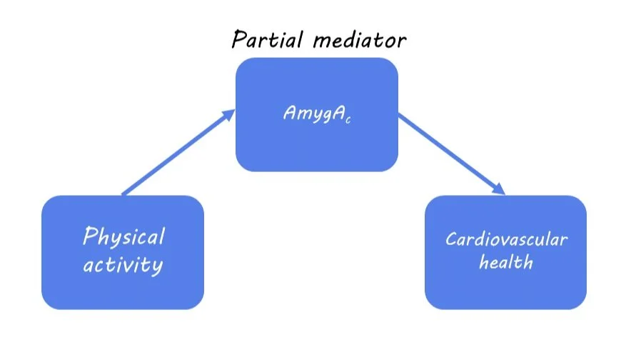
The health questionnaires provided information on the history of cardiovascular events (such as heart failure or stroke) and psychiatric disorders (such as depression). The authors used regressions to statistically assess relationships among the variables. First, they examined the associations between physical activity and AmygAc. Then they assessed the association between physical activity and cardiovascular events. Finally, they assessed the association between AmygAc and cardiovascular events. With these sets of equations, they were able to assess whether AmygAc mediated the relationship between physical activity and cardiovascular events. That is to say, whether physical activity indirectly impacts cardiovascular health via stress reduction in the brain’s stress pathways. History of depression was also included as an interaction variable in these models to allow the authors to understand whether patterns in the relationships among the other variables were different for people with past depression.

What did they find?

First, the authors found that physical activity was associated with a decrease in stress-related neural activity, such that the more physical activity participants reported, the less stress-related activity they showed. This was, in large part, associated with increases in prefrontal cortical activity, which may contribute to cognitive health benefits. Second, as expected, they found that physical activity was associated with decreased risk of cardiovascular events. Third, they found that increased AmygAc, representing increased stress-related activity, was associated with an increased risk of cardiovascular events. These three findings aligned well with their hypotheses: 1) more physical activity is related to less stress activity in the brain, 2) more physical activity is related to less risk of cardiovascular events, and 3) more stress-related activity is related to more risk of cardiovascular events.

Next, they evaluated whether physical activity lowers cardiovascular disease risk by reducing AmygAc. From their mediation analysis, the authors found that AmygAc was a partial mediator of the relationship between physical activity and cardiovascular events. This means that one way in which physical activity is associated with improved cardiovascular health is by reducing stress, but it’s not the only underlying mechanism.

Finally, the authors found that the benefits of physical activity on cardiovascular health were highest among people with a history of depression. Specifically, people without a history of depression experienced the anticipated benefits of physical exercise, and their risk reductions plateaued at ~300 minutes of moderate-intensity activity per week. On the other hand, people with a history of depression derived roughly double the overall cardiovascular risk reductions compared to those without depression, and continued to derive benefits at the higher levels of activity.

What's the impact?

In this paper, the authors demonstrate that physical activity reduces stress-related brain activity and the risk of cardiovascular events, especially for people with a history of depression. Importantly, reduced stress partially mediates the relationship between physical activity and cardiovascular events, providing an indication of the underlying mechanisms. Future work may further characterize other mediating factors among these variables.

A Newly Identified Body-to-Brain Circuit for Regulating Inflammation

Post by Shahin Khodaei

The takeaway

Communication between the brain and the body plays a key role in the immune response: information about bodily inflammation is delivered to the brain, which can subsequently adjust inflammation in the body to an appropriate level.

What's the science?

Inflammation, a component of innate immunity, is an important part of the body’s response to damage or pathogens. Several studies have shown that the brain can detect the state of inflammation in the body and that neurons can modulate inflammation through their activity. However, the pathways by which the brain regulates bodily inflammation are still not well understood. This week in Nature, Jin and colleagues published a study that identified: 1) specific populations of neurons that carry information about the state of inflammation in the body to the brain, and 2) specific populations of neurons in the brain that respond to this information to ramp up or dampen inflammation in the body.

How did they do it?

To cause inflammation in mice, the authors injected them with lipopolysaccharide (LPS) – a component of bacterial cell walls that is recognized by immune cells as a signal for pathogens. In response, these cells release protein messengers known as cytokines, which can be pro-inflammatory (heighten inflammation) or anti-inflammatory (reduce inflammation). 

The authors then had two major goals: to determine which neurons respond to inflammation and to determine how the activity of these neurons shapes the inflammatory response.

For their first goal, the authors used imaging techniques to find out which neurons are activated after LPS, and pro- or anti-inflammatory cytokines. They then used single-cell RNA sequencing to map the genes that are expressed by these neurons, to find markers to identify them.

To manipulate the activity of neurons, the authors used the genetic technology of designer receptors exclusively activated by designer drugs or DREADDs. Depending on the type of DREADD (excitatory vs. inhibitory) inserted into cells, the authors can selectively increase or decrease the activity of sub-populations of neurons. In conjunction, the authors used another genetic technology called TRAP (targeted recombination in active populations), which allowed them to express DREADDs only in neurons that were involved in inflammation. This approach lets them manipulate the activity of inflammation-responsive neurons.

What did they find?

Using their imaging techniques, the authors found a population of neurons in the brainstem that was activated by LPS-triggered inflammation. These neurons received information about bodily inflammation from the vagus nerve. The authors found two distinct populations of vagal neurons: one responding to pro-inflammatory cytokines and expressing the genetic marker TRPA1, and the other to anti-inflammatory cytokines and expressing the genetic marker CALCA. In this way, vagal neurons transmit information regarding the inflammatory state to the brainstem.

How does manipulating the activity of these neurons affect the inflammatory response? Using DREADDs, increasing the activity of brainstem neurons was anti-inflammatory, while inhibiting their activity during LPS injection led to much higher inflammation. Similarly in the vagus nerve, increasing the activity of either the TRPA1 or the CALCA subpopulations had an anti-inflammatory effect. Remarkably, the authors further showed that in a mouse model of deadly inflammation, activating this body-brain circuit using DREADDs significantly reduced the likelihood of death.

What's the impact?

This study identified a specific body-to-brain circuit that regulates inflammation. By monitoring the levels of pro- and anti-inflammatory cytokines in the body, this circuit monitors the inflammatory response and regulates inflammation levels as needed. In the future, targeting this system may provide new strategies for treating diseases that involve dysregulation of the inflammatory response.

Access the original scientific publication here. 

Investigating the Organization of Brain Tumor Cells Using New High-Resolution Technologies

Post by Natalia Ladyka-Wojcik

The takeaway

Using new technologies for studying the spatial architecture of gliomas reveals both local and global organization that are largely driven by hypoxia (i.e., when oxygen is not sufficiently available at the tissue level), providing critical insights for the future of cancer treatment research.

What's the science?

Glioma is a type of cancer that starts as a growth of cells in the brain or spinal cord, rapidly invading and destroying healthy surrounding tissue. Critically, gliomas are characterized by a very complex spatial architecture, making it difficult to determine the organization of their cell types and cellular states. Until recently, histopathology – or the examination of cancerous tissue under a microscope – was the dominant method for studying cell types and cellular states of gliomas, but histopathology lacks the granularity to fully capture the spatial architecture of gliomas. New technological developments have been made in spatial transcriptomics, a molecular profiling method allowing researchers to measure all gene activity in a tissue sample. When paired with advances in the study of proteins (i.e., proteomics), researchers are enabled to measure all gene activity in a tissue sample, offering new opportunities to map the complex spatial architecture of gliomas. This week in Cell, Greenwald and colleagues profiled glioma tissue samples using these new technologies to develop a framework for systematically describing the spatial organization of gliomas.

How did they do it?

The authors investigated glioma samples from patients who had undergone tumor resection across multiple hospital sights, and whose tumors ranged in their specific location in the brain as well as in their key biomarkers. These samples were frozen by liquid nitrogen for preservation and then profiled using spatial transcriptomics within one week. Broadly, the goal of spatial transcriptomics is to count the number of transcripts of a gene at distinct spatial locations in a tissue. More specifically, the authors used a commercialized transcriptomics technique, called “Visium”, to spatially profile the glioma samples at a high level of spatial granularity. This allowed the authors to investigate not only the patterns of organization across gliomas but also to determine to what degree the spatial location of gliomas affects the diversity of cellular states.

What did they find?

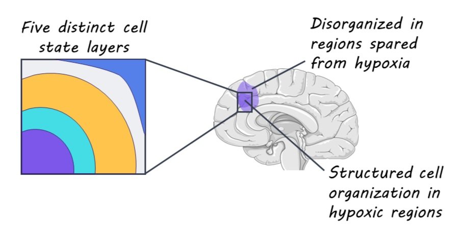
The authors identified three key modes of glioma organization, each respectively focused on 1) the local environment of glioblastoma tumors, 2) the pairing of cellular states across tumors, and 3) the global architecture of the tumors. The first key mode of glioma organization that the authors found is that cells tend to be surrounded by other cells in the same state, forming local environments that are highly homogeneous in configuration and gene expression. This finding suggests that spatial location plays an important role in the regulation of the cell state. The second key mode of glioma organization that the authors reported had to do with how pairs of states are arranged across multiple scales. That is, pairs of cellular states or gene expression patterns tend to be consistently associated with each other across different scales within the tumor tissue. Importantly, understanding these state-to-state associations across different spatial scales can help us to better understand the developmental processes of gliomas. Finally, the third key mode of glioma organization that the authors found is related to global arrangement of tissue layers. Specifically, the authors detected five distinct layers, with cell states in each layer being associated with the same layer or adjacent ones. Critically, the authors discuss that hypoxia might drive the organizational characteristics of glioma tumors such that regions spared from hypoxia are actually relatively disorganized in comparison.

What's the impact?

This study is the first to characterize both local and global organizational features of glioma tumors at a highly granular level using new advances in spatial transcriptomics and proteomics. The three key organizational modes identified in this study provide critical insights into how hypoxia drives the spatial architecture of gliomas, which in turn can support the development of targeted treatments for glioblastoma.  

Access the original scientific publication here.