How Brain Connectivity Contributes to Different Types of Goal Pursuit

Post by Lila Metko

The takeaway

There is lower connectivity between movement-related brain regions in individuals with a higher propensity to use the ‘prevention system’ to pursue goals that prevent a negative outcome rather than the ‘promotion system’ to pursue goals that have a positive outcome.

What's the science?

According to regulatory focus theory, there are two major cognitive-motivational systems involved in accomplishing goals: the promotion system, involved in achieving hopes and dreams, and the prevention system, involved in fulfilling duties and obligations. The promotion system is oriented towards making good things happen, while the prevention system is focused on preventing negative things from happening. Individual differences in these systems of self-regulation have been linked to psychopathology. There are previously established brain regions that have been shown to be involved in each of these systems and they share some overlap. This week in PNAS Nexus, Kim and colleagues used functional magnetic resonance imaging (fMRI) data to create a predictive network model capable of predicting differences in propensity towards regulatory focus systems based on connectivity between neural structures. 

How did they do it?

The authors studied 1,307 university students enrolled in the Duke Neurogenetics Study. They gave the participants the Adolescent Regulatory Focus Questionnaire (RFQ), a questionnaire that measured their inclination towards a promotion system or prevention system way of attaining goals. fMRI scans were taken of the participants in both resting state (patients awake with eyes open but with no task) and during emotional face matching, card guessing, working memory, and face naming tasks. The authors then estimated general functional connectivity for each participant by combining resting state and task fMRI data and regressing out task-related events, to obtain more reliable data than if they used resting fMRI alone. A predictive model of regulatory focus orientation was created using correlations from participants’ functional connectivity to their scores in the RFQ. A group of participants was left out of the creation of the model to test its ability to predict regulatory focus orientation. To test this, they compared actual scores to predicted scores in the group that was left out. 

What did they find?

The model generated by the authors was predictive of prevention but not promotion scores. It’s possible the lack of prediction for promotion scores is because promotion-orientated processes do not require as complex cognitive processes as prevention and thus may not be detectable in these measures of functional connectivity. It was found that lower functional connectivity between association cortices (brain regions involved in understanding sensory information and planning a behavioral response) was correlated with increased prevention. It had been previously established that these association regions were important for prevention behaviors, but interestingly the authors found a novel region associated with prevention; the primary motor cortex. More than half of the measures of brain connectivity that had a negative correlation with prevention scores involved the primary motor cortex.  

What's the impact?

This study is the first to show that the primary motor cortex, a region involved in initiating voluntary movements, contributes to prevention system function. It is also the first study to create a predictive model for these types of preventative behaviors in a large sample. Importantly, regulatory focus orientations are predictive of vulnerability to psychopathology and improper function of these regulatory systems may also be predictive of generalized anxiety disorder and depression. Thus, the study of brain regions involved in regulatory focus is of high clinical significance.  

Access the original scientific publication here.

REM Sleep Protects Against Fearful Memories

Post by Meagan Marks

The takeaway

REM sleep may protect against the development of post-traumatic stress disorder by enhancing the brain’s ability to extinguish fearful memories. It does so by strengthening the excitability of infralimbic cortex neurons, which play a crucial role in fear extinction. 

What's the science?

Post-traumatic stress disorder (PTSD) often presents as persistent, uncontrollable fear responses triggered by cues associated with a past traumatic event. PTSD is likely caused by a neural disruption in fear extinction, which is the process of learning that a fear-inducing cue is no longer predictive of danger. Fear extinction is carried out by neurons in the infralimbic cortex (IL) in mice, which is homologous to the ventromedial prefrontal cortex in humans. It is a region highly active during rapid eye movement (REM) sleep. Interestingly, PTSD patients often experience disturbances in REM sleep, but the exact role that it plays in fear extinction remains unknown. This week in Current Biology, Hong and colleagues determine how REM sleep influences fear extinction, particularly through its influence over IL neuron excitability.

How did they do it?

To understand the paired role that REM sleep and IL neurons play in fear extinction, the authors conducted an experiment over the course of three days. On the first day, the mice were fear conditioned, learning that an auditory cue (20-second tone) would elicit an unpleasant stimulus (one-second foot shock). Immediately after this conditioning, the authors used optogenetics to silence the activity of IL neurons during sleep. At this stage, the mice were split into three groups: for one group, IL neurons were silenced only during REM sleep, while for another, the neurons were silenced immediately after REM sleep. A control group with no neural manipulation was also included. 

On the second day, the mice were placed back in the fear conditioning environment, but this time, the auditory cue was played without a subsequent foot shock. This was a day of extinction learning, where the mice learned that the auditory cue should no longer induce fear. 

On the third and final day, the auditory cue again played with no subsequent foot shock. This was a day of recall, where the authors observed how well the mice extinguished the fear association from the day before. They did so by measuring how long the mice froze after each auditory cue, an innate fear response in rodents. 

Additionally, the authors repeated this paradigm twice more, with slight changes. During one repeat, the authors waited an additional four hours after conditioning to silence IL neurons during REM sleep. During the second, IL neurons were not silenced after fear conditioning but instead were immediately silenced after extinction learning.

What did they find?

The authors found that inhibiting IL activity during REM sleep post-conditioning significantly increased freezing time during recall. The control mice and mice with inhibition after REM sleep did not freeze as much, suggesting that IL activity during REM sleep is crucial to the consolidation of fear extinction memories. Delaying the post-conditioning inhibition of IL activity during REM sleep did not significantly alter freezing, nor did it inhibit IL neurons immediately after extinction learning.

Looking at these findings altogether, it can be concluded that IL neuron activity during REM sleep that occurs immediately after fear conditioning is crucial to fear extinction. This is because inhibiting IL neuron activity decreases the overall excitability of the neurons, and therefore, inhibiting IL neuron activity during all of REM sleep when the neurons are most active decreases their excitability the following day. This hindered their ability to fire during extinction learning, and the mice were less capable of encoding new fear extinction memories. 

What's the impact?

This study found that REM sleep plays a crucial role in fear extinction by strengthening the excitability of IL neurons and enhancing their ability to encode new extinction memories. This is an important finding when it comes to PTSD, as many patients struggle with disturbances in REM sleep and experience reduced activity in extinction-encoding neurons. Knowing that REM sleep plays a pivotal role in extinction memory offers great potential in finding new therapies for PTSD and gives greater insight into the circuitry of fear extinction. 

Access the original scientific publication here

Understanding Compulsive Disorders

Post by Laura Maile

What is compulsion?

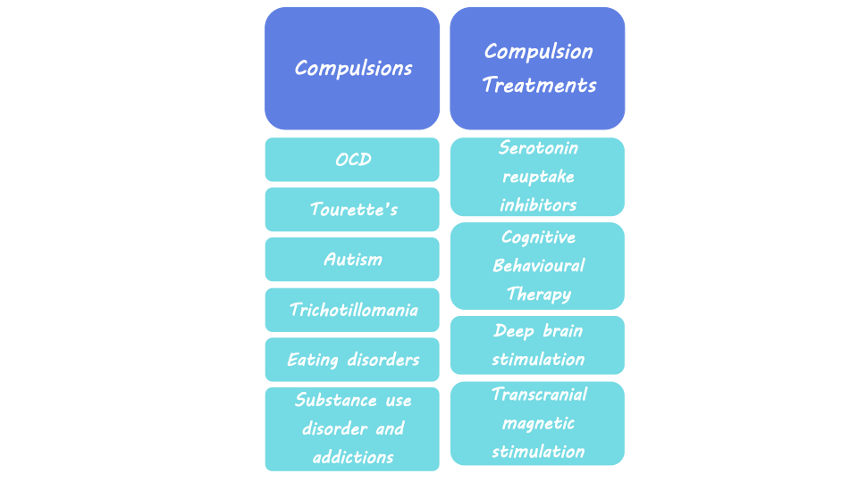
Compulsions are described as persistent urges to perform a specific behavior, even when the consequences are negative. They induce a feeling that the individual must complete the behavior, even though it may conflict with their overall goal. These repetitive behaviors are maladaptive, meaning they prevent individuals from adapting to their environment and are detrimental to overall well-being. Compulsions are a symptom of diseases like Obsessive Compulsive Disorder (OCD) and Tourette’s Syndrome, but they can occur in many other psychiatric disorders including Autism Spectrum Disorder, trichotillomania, eating disorders, substance use disorder, and other addictions. While compulsivity is a maladaptive symptom of disease, it can also be a component of everyday human behavior that leads to positive outcomes, including behaviors such as proofreading or athletic performance rituals.  

Neurological basis of compulsive behavior

Some theories of compulsive behavior, which generally include ideas of positive and negative reinforcement learning, suggest that the neural networks that control goal-directed behavior and habits are out of balance. These networks include brain regions important for goal-directed and habitual behavior like the striatum, a part of the basal ganglia, which is important for controlling motor behavior, learning, and decision-making. In rodent studies where drugs are administered to inactivate specific areas of the striatum, habitual seeking of food and alcohol is reduced. Rodent studies also show that compulsive behavior can be born out of negative reinforcement, or avoidance of negative outcomes. The emergence of this compulsivity is dependent on brain systems required for habit formation like the striatum. The medial prefrontal cortex (mPFC) is also important for goal-directed behavior, habit formation, behavioral flexibility, the association between actions and outcomes, and decision-making. The nearby orbitofrontal cortex (OFC), a region involved in decision-making related to reward and emotion, is also likely involved in compulsive behavior. Another region implicated in goal-seeking and habitual behavior is the amygdala, which is part of the limbic system associated with emotion, fear, and aspects of learning and memory that are influenced by emotionally important stimuli.  

Compulsive behavior can result when there is an imbalance or impairment in these networks that regulate goal-directed behavior. Neuroimaging studies in the human brain suggest that changes in these brain regions that regulate behavior are related to compulsion. For example, studies show reduced brain volume (i.e., grey matter) and elevated brain activity in the OFC in individuals with OCD. Changes in neural connectivity between the prefrontal cortex and striatum are also seen in patients with OCD.  

Human imaging studies of individuals with OCD and substance use disorder found reduced functional connectivity (i.e., neural synchrony between brain regions) and activity of prefrontal cortex regions, which correlated with compulsive behavior. This loss of connectivity in the regions governing goal-directed behavior suggests a loss of control over habitual behavior and an inability to inhibit future actions, leading to compulsivity. Changes in activity related to compulsivity have also been seen in the insula, a region of the cortex associated with interoception, or awareness of bodily feelings. Interoceptive feelings and insula activity may trigger habits that have been developed through experiences involving negative reinforcement. The variety of findings in human imaging studies demonstrates the complexity of these neural networks that interact to influence compulsive behavior and highlight the need for more detailed research.

Treating compulsive behavior

Treatments for OCD and compulsive behaviors associated with other psychiatric disorders include serotonin reuptake inhibitors, cognitive behavioral therapy, and deep brain stimulation.  Deep brain stimulation includes the implantation of electrodes into the brain to allow for electrical stimulation of specific areas. Clinical studies have shown that deep brain stimulation of the nucleus accumbens, a region associated with reward, was successful at reducing compulsivity symptoms in OCD patients who did not respond to other forms of treatment. A less invasive form of treatment is transcranial magnetic stimulation, which uses a magnetic coil outside the skull to influence the activity of certain brain areas. Transcranial magnetic stimulation of the mPFC has been shown to reduce OCD symptoms in some patients.  Other studies using this technology to stimulate regions like the OFC, striatum, and basal ganglia show some promise for treating intractable OCD, though results are mixed.  Transcranial magnetic stimulation has also been successfully deployed to target regions of the prefrontal cortex to reduce compulsive drug seeking in patients with substance use disorder.  

What's next?

While some success has been achieved with transcranial and deep brain stimulation, more research is needed to improve outcomes and reduce side effects of compulsive behavior. New deep brain stimulation techniques are being investigated that can deliver neurostimulation while recording neural activity, which will allow scientists more insight into the neural basis of compulsive behavior. Other novel treatments being studied for compulsive behavior include ketamine and psychedelics, which can be used as treatments for depression or anxiety. There is also ongoing work aimed at uncovering how findings related to the contribution of serotonin, dopamine, and noradrenaline to compulsive behavior translate from rodent to human systems.  

References +

Robbins et al. From compulsivity to compulsion: the neural basis of compulsive disorders. Nat. Rev. Neurosci. (2024).

Robbins et al. Obsessive-Compulsive Disorder: Puzzles and Prospects. Neuron (2019).

Marti-Pratz et al. The development of compulsive coping behavior depends on dorsolateral striatum dopamine-dependent mechanisms. Nature Portfolio (2023).

Simmler et al. Neural circuits in goal-directed and habitual behavior: Implications for circuit dysfunction in obsessive-compulsive disorder. Neurochemistry International (2019).

Anticevic et al. Global resting-state functional magnetic resonance imaging analysis identifies frontal cortex, striatal, and cerebellar dysconnectivity in obsessive-compulsive disorder. Biol. Psychiatry (2014).

Denys et al. Deep Brain Stimulation of the Nucleus Accumbens for Treatment-Refractory Obsessive-Compulsive Disorder. JAMA Psychiatry (2010).

Goodman et al. Harmonizing the Neurobiology and Treatment of Obsessive-Compulsive Disorder. American Journal of Psychiatry (2021).