Microglial Cell Memory Can Change Neuropathology

What's the science?

Microglia are the resident macrophages of the brain’s innate immune system and respond to injury or pathogens. Some innate immune cells in the body have a memory; they may exhibit a ‘training response’, meaning they show an increased inflammatory response to a pathogen the second time it is presented. They may also develop another type of memory called ‘tolerance’ where inflammation is reduced after a pathogen is presented numerous times. We don’t know whether immune training or tolerance exist in microglia or whether these features play a role in shaping neurological diseases later in life. This week in Nature, Wendeln and colleagues explored whether microglial-mediated immune responses in the brain depend on the history of immune responses, indicating immune memory.

How did they do it?

First, they tested whether immune activation in the body (periphery) induced immune memory or tolerance in the brain. Control mice and microglia knockout mice were given lipopolysaccharides (LPS), an inflammatory molecule that induces sickness behavior and peripheral inflammation, up to four times, and the immune response was observed after each administration. Next, to test whether immune memory or tolerance affects neuropathology, they used two models: First, they used APP23 mice, in which amyloid-β plaques are produced (Alzheimer’s disease pathology model). Second, they used an ischemia model (inducing brain ischemia as a model for stroke). They tested whether peripheral LPS stimulation induced immune memory in the brain and modulated later occurring neuropathologies. Finally, they isolated microglia from mice at 9 months of age who had been treated with one or four LPS administrations 6 months prior, and looked at markers for enhancers (regulatory elements of DNA that enhance expression of certain genes) to understand whether epigenetic factors underlie the microglia responses.

What did they find?

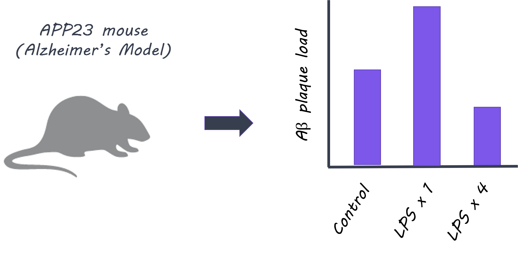
When LPS was administered to control mice, more cytokines (normally released as part of an inflammatory response) were released in the brain after the second administration compared to the first, indicating that ‘immune training’ in the brain does occur. This response was not seen in microglia knockout mice, demonstrating that microglia play a key role in immune training. In contrast, cytokine release in the brain diminished after four LPS injections (i.e. a larger number of exposures to peripheral inflammation), indicating ‘immune tolerance’ in the brain. In the next experiment, APP23 mice (Alzheimer’s pathology model) were examined 6 months after LPS treatment (applied before brain pathology developed). Here, brain plaques were increased in mice who had been administered LPS once (suggesting immune training could increase plaque occurrence), and decreased in mice who had been treated with four LPS injections (suggesting immune tolerance could reduce plaques). Treatment with LPS also altered the brain’s immune response to plaque deposition, as shown by changes in certain cytokine levels. In the ischemia (stroke) model, mice administered LPS once showed increased levels of cytokines in the brain, while mice administered LPS four times, showed decreased levels, demonstrating immune training and tolerance respectively. Brain damage following ischemia was reduced only in mice administered LPS four times, indicating that immune ‘tolerance’ may be protective against future neuropathology. In isolated microglia, markers for enhancers were increased in different signaling pathways after one LPS administration (immune ‘training’ response) versus after four administrations (immune ‘tolerance’ response), indicating that epigenetic changes in microglia following peripheral immune stimulation underlie these long-term effects.

Alzheimer’s and amyloid-beta plaques

What's the impact?

This is the first study to characterize the ‘memory’ of the innate immune response of microglia in the brain and its role in modifying neuropathologies. The results suggest that certain neuropathologies (such as Alzheimer’s or stroke) may be altered by microglial immune memory due to much earlier occurring immune stimulation in the periphery. Next, it will be important to understand precisely which immune stimuli change the microglial response and in what way.

A. Wendeln et al., Innate immune memory in the brain shapes neurological disease hallmarks. Nature (2018). Access the original scientific publication here.

The Brain’s Immune Cells Help Motor Neurons Recover in a Mouse Model of ALS

What's the science?

ALS is a devastating disease where motor neurons degenerate. TDP-43 is a toxic protein that builds up in ALS and contributes to this degeneration. It is unclear whether the immune cells of the nervous system, microglia, react to TDP-43 and whether they play a protective role or make neurodegeneration worse. This week in Nature Neuroscience, Spiller and colleagues test whether microglia contribute to neurodegeneration in a mouse model of ALS.

How did they do it?

They used a transgenic mouse model for ALS in which TDP-43 (toxic protein) build-up can be induced or reversed by suppressing or activating TDP-43 gene expression. With this model they can simulate motor neuron degeneration or recovery. The authors measured whether there were any changes in the number of microglia or their activation state (indicated by the size and shape of the cells) at several time points using immunostaining in the spinal cord, both after inducing TDP-43 damage and after reversing this process. Increases in microglia number and activation indicate that the microglia are actively responding to neuron damage. They also measured gene expression changes in the microglia during the degeneration and recovery of motor neurons. They then measured whether TDP-43 could be found inside the microglial cells, indicating that the microglia are “eating” the proteins to clear them. Finally, they blocked microglia cell replication to test how motor neuron function would recover in regenerating motor neurons.

What did they find?

Microglia were not initially reactive to toxic protein buildup. By the time the motor neurons were severely degenerated, there was a slight increase in the number of microglia and a change in the activation state (i.e. the number of microglia reacting). After reversing the TDP-43 pathology, microglia number and activation increased immediately and dramatically, suggesting that the microglia play a more important role in helping motor neurons recover. This activation of microglia was specific to TDP-43 proteins, and not just a response to neuron death in general. The microglia also showed many changes in gene expression during early recovery (and not early disease), indicating that they are involved in the early recovery process. TDP-43 was found inside microglia, showing that these immune cells actually clear TDP-43 during recovery. When microglial cells were not able to multiply, the motor neurons that were damaged by TDP-43 did not fully recover, and the mouse did not regain full motor function, indicating that microglia are required for a full motor recovery.

Microglia, Servier Medical Art, image by BrainPost, CC BY-SA 3.0

Microglia, Servier Medical Art, image by BrainPost, CC BY-SA 3.0

What's the impact?

This is the first study to show that microglia are involved in the recovery of motor neurons in a reversible mouse model of ALS. Previously, we weren’t sure whether these immune cells of the nervous system actually make things worse or protect neurons from damage in ALS. We now know that microglia play an active role in restoring motor function in neurons damaged by TDP-43 protein buildup in this mouse model of ALS. Currently, we do not have therapies to restore motor function in ALS, and this study suggests that we may be able to target microglia.

krista_test_small.png

Reach out to study author Dr. Krista Spiller on Twitter @krista_spiller

K. Spiller et al., Microglia-mediated recovery from ALS-relevant motor neuron degeneration in a mouse model of TDP-43 proteinopathy. Nature Neuroscience (2018). Access the original scientific publication here.

Astrocytes Become Reactive with Normal Aging

What's the science?

Astrocytes are are the most abundant cell in the brain. They help to respond to injury and are important for maintaining overall brain health by supporting neurons, recycling neurotransmitters and regulating the formation and elimination of the connections between neurons. Astrocyte dysfunction is known to play a role in neurodegenerative diseases, but how astrocytes change throughout normal aging is not well known. One way to understand these changes is by looking at the transcription of genes in astrocytes. This week in PNAS, Clarke and colleagues performed RNA sequencing in mice at different stages of life to understand how astrocytes change over time.

How did they do it?

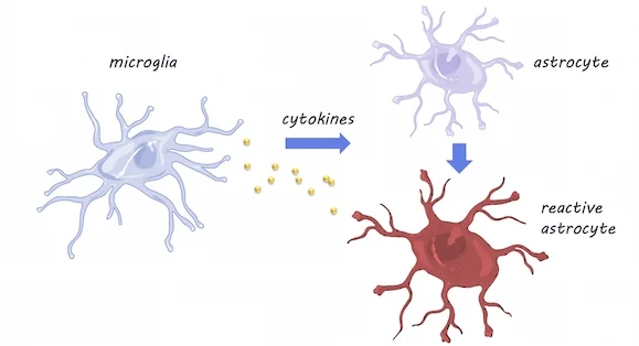
RNA sequencing was performed in mice at five time points between adolescence and old age, in three different brain areas: the cortex, hippocampus (involved in memory), and striatum (involved in movement and reward). They validated their findings using fluorescence in situ hybridization and quantitative polymerase chain reaction (qPCR) techniques (these techniques can confirm gene expression changes). To investigate whether the resident immune cells of the brain - microglia - play a role in inducing changes in astrocytes with aging, they compared astrocyte gene expression in mice with and without (knock-out mice) cytokines. Cytokines are released by microglia in response to neuroinflammation. 

What did they find?

Using RNA sequencing, they found that as astrocytes age, they are more likely to express genes associated with reactivity (this is when astrocytes become dysfunctional -- typically associated with neuroinflammation). Astrocytes were especially likely to become reactive in the hippocampus and striatum, which are areas particularly susceptible to neurodegeneration in aging. Using qPCR, a method used to observe DNA sequences, they found that reactive gene expression was not increased in the knock-out mice without cytokines, indicating that microglia expression of cytokines may be partially responsible for changes in astrocyte gene expression. Aged brains also formed many more reactive astrocytes in response to the neuroinflammation inducer ‘lipopolysaccharide’, which may indicate vulnerability of the aged brain to disease and inflammation.

                       Microglia & Astrocytes, Servier Medical Art, image by BrainPost, CC BY-SA 3.0

                       Microglia & Astrocytes, Servier Medical Art, image by BrainPost, CC BY-SA 3.0

What's the impact?

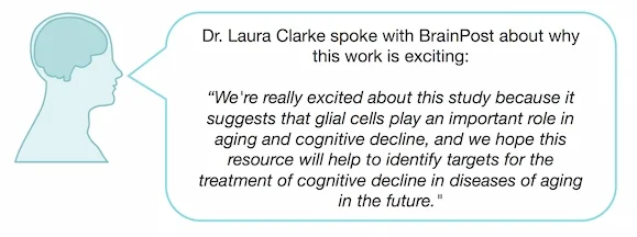
This is the first study to demonstrate that astrocytes become reactive as they age and that microglia- the immune cells of the brain- may be responsible through cytokine activity. More reactive astrocytes were found in brain regions vulnerable to degeneration, suggesting that changes in astrocyte gene expression may help explain neurodegenerative diseases or cognitive decline in aging.

Clarke_comment_new_small.jpg

Reach out to study author Dr. Laura E. Clarke on Twitter@ClarkeLauraE

Clarke et al., Normal aging induces A1-like astrocyte reactivity. (2018). Access the original scientific publication here.