Different Profiles of Microglial Activation in Alzheimer's disease

What's the science?

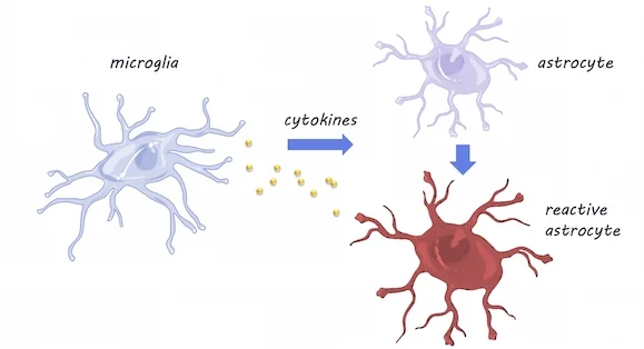
Microglia, the immune cells of the brain, may contribute to Alzheimer’s disease by becoming activated in response to brain pathology (also known as neuroinflammation). Currently, whether neuroinflammation is associated with Alzheimer’s progression (harms the brain) or whether it may be protective (helps to “eat” plaques in the brain) is still a matter of debate. This week in BrainHamelin and colleagues used PET imaging to examine how microglial activation in the brain is related to Alzheimer’s disease progression.

How did they do it?

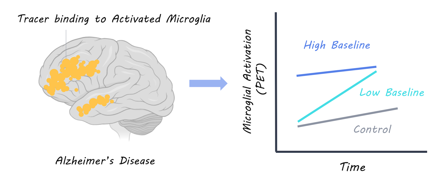
PET imaging was used to measure the uptake of a radiotracer (18F-DPA-714) in the brain binding to activated microglia. A large group of patients with Alzheimer’s disease were scanned twice for activated microglia, once at baseline and once two years later. They were then followed up annually and scanned with MRI to measure brain volume (measure of Alzheimer’s progression) and given annual cognitive tests to assess dementia severity and cognitive function. Based on this, patients were split into “fast and slow decliner” categories. The microglial activation levels over time were also analyzed compared to a control group of healthy participants.

What did they find?

Having a high level of microglial activation at baseline was predictive of being a slow decliner. In patients with a high baseline neuroinflammation, cognitive performance was better and brain volume was more preserved, suggesting that more microglial activation at baseline is protective. At two years follow-up, microglial activation was higher in Alzheimer’s participants but not controls as would be expected. Increased microglial activation over time was related to worsening cognitive scores and brain atrophy, suggesting that it is harmful. However, when they examined neuroinflammation over time at an individual level, they found that those with the highest baseline microglial activation had the lowest increase in microglial activation over time. They concluded that there is a dynamic relationship, whereby neuroinflammation may affect patients differently, depending on their original level of microglial activity. Microglial activation appears to be protective initially, but exacerbates Alzheimer’s disease over time; to a greater extent in those who had low levels of microglial activation to begin with.

Microglial activation in Alzheimer’s disease

What's the impact?

This is the first study to show that neuroinflammation may affect individuals with Alzheimer’s disease differently depending on their baseline level of microglial activity. It shows us that microglial activation may be helpful or harmful depending on the individual and how far their disease has progressed. Understanding the role microglial activation play in Alzheimer’s disease is an essential part of understanding how the disease progresses.


L. Hamelin et al., Distinct dynamic profiles of microglial activation are associated with progression of Alzheimer's disease. Brain (2018). Access the original scientific publication here.

Astrocytes Become Reactive with Normal Aging

What's the science?

Astrocytes are are the most abundant cell in the brain. They help to respond to injury and are important for maintaining overall brain health by supporting neurons, recycling neurotransmitters and regulating the formation and elimination of the connections between neurons. Astrocyte dysfunction is known to play a role in neurodegenerative diseases, but how astrocytes change throughout normal aging is not well known. One way to understand these changes is by looking at the transcription of genes in astrocytes. This week in PNAS, Clarke and colleagues performed RNA sequencing in mice at different stages of life to understand how astrocytes change over time.

How did they do it?

RNA sequencing was performed in mice at five time points between adolescence and old age, in three different brain areas: the cortex, hippocampus (involved in memory), and striatum (involved in movement and reward). They validated their findings using fluorescence in situ hybridization and quantitative polymerase chain reaction (qPCR) techniques (these techniques can confirm gene expression changes). To investigate whether the resident immune cells of the brain - microglia - play a role in inducing changes in astrocytes with aging, they compared astrocyte gene expression in mice with and without (knock-out mice) cytokines. Cytokines are released by microglia in response to neuroinflammation. 

What did they find?

Using RNA sequencing, they found that as astrocytes age, they are more likely to express genes associated with reactivity (this is when astrocytes become dysfunctional -- typically associated with neuroinflammation). Astrocytes were especially likely to become reactive in the hippocampus and striatum, which are areas particularly susceptible to neurodegeneration in aging. Using qPCR, a method used to observe DNA sequences, they found that reactive gene expression was not increased in the knock-out mice without cytokines, indicating that microglia expression of cytokines may be partially responsible for changes in astrocyte gene expression. Aged brains also formed many more reactive astrocytes in response to the neuroinflammation inducer ‘lipopolysaccharide’, which may indicate vulnerability of the aged brain to disease and inflammation.

                       Microglia & Astrocytes, Servier Medical Art, image by BrainPost, CC BY-SA 3.0

                       Microglia & Astrocytes, Servier Medical Art, image by BrainPost, CC BY-SA 3.0

What's the impact?

This is the first study to demonstrate that astrocytes become reactive as they age and that microglia- the immune cells of the brain- may be responsible through cytokine activity. More reactive astrocytes were found in brain regions vulnerable to degeneration, suggesting that changes in astrocyte gene expression may help explain neurodegenerative diseases or cognitive decline in aging.

Clarke_comment_new_small.jpg

Reach out to study author Dr. Laura E. Clarke on Twitter@ClarkeLauraE

Clarke et al., Normal aging induces A1-like astrocyte reactivity. (2018). Access the original scientific publication here.