Modeling Sound Localization in the Brain

Post by Anastasia Sares

The takeaway

Tiny delays in sound between our ears help us determine the location of sounds in space. Using simulations of neuronal activity, the authors compared two models of auditory delay processing and found that a model supported by research in mammals better accounted for auditory delay processing than a model supported by research in other animals (mainly birds).

What's the science?

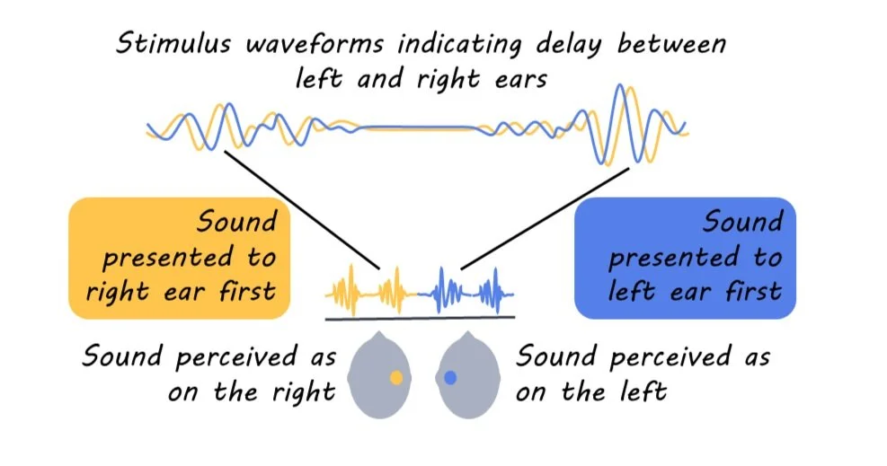
We don’t often think about it, but our auditory system is extremely good at localizing sound. This is what makes it possible to hear a car driving up behind you, or to automatically reach for your phone when it buzzes. Among the main cues that help us localize sound are inter-aural timing differences (ITDs), where sound arrives at one ear slightly before the other. The differences can be minute (on the order of millionths of a second) and are calculated within the first few synapses of the auditory system—which are extremely difficult to probe and can only be accessed with invasive surgery. Because of this, even though we have known about the importance of these timing cues since at least the 1800s, we still don’t have a complete idea of how they are represented in the human brain. Many psychology courses still teach the Jeffress model, which assumes that the calculation of ITDs is mostly due to differing lengths of axons coming in from either side of the head. This model finds some support in animal research (like in owls), but it is not certain that primates’ auditory systems work the same way.

This week in Current Biology, Undurraga and colleagues combined neuroimaging data and computer simulations to show how inter-aural time delays might be processed in humans.

How did they do it?

Participants’ electroencephalography (EEG) and magnetoencephalography (MEG) data were recorded while they listened to a noise stimulus through headphones. The noise varied in inter-aural timing difference (ITD) between the two ears. Sometimes, the sound from the left ear was played slightly earlier (simulating a sound coming from the left) and sometimes, the sound from the right ear was played slightly earlier (simulating a sound coming from the right), and there were varied amounts of delay on different trials. The experiment included realistic delay for direct sound coming from a nearby object (500 microseconds) as well as much longer delays that might indicate reverberations from a far-away source (up to 4000 microseconds).

The authors then looked for fluctuations in the brain data that matched the rate of changes in ITD while being insensitive to other changes in the signal (like intensity/loudness or frequency/pitch). These fluctuations in the brain data that are locked to the changing ITD are termed the ITD-following response.

Finally, the authors created simulations of neuronal activity based on different theories about how ITD is processed, one closer to Jeffress’ delay-line model (with more support in bird studies), and one called the π-limit model (with some support in other mammals). They measured how well these simulated neuronal arrays could reproduce the human data.

What did they find?

The ITD-following response appeared in multiple brain areas. Unsurprisingly, the auditory cortex was highly responsive to changes in ITD, but somatosensory regions were also fairly responsive. The right hemisphere responded more than the left. The ITD-following response was strongest at the shortest delay (500 microseconds). However, the response didn’t just fall off at longer delays, there was a strange oscillating pattern, indicating that some delays provoked a bigger response than others.

Simulations indicated that the mammal-like model was more appropriate than the bird-like model in explaining the human brain activity data. In fact, when fitting the bird model to human data, it ended up taking on the properties of the mammal-like model anyway. As for the strange oscillating pattern at very long delays, the authors found it was likely a side-effect: a convergence of responses from neurons that primarily respond to shorter ITDs. The oscillations were sort of like “echoes” of the short-delay neurons’ activity, stretching out to a longer time frame. This means there is probably not a separate dedicated mechanism producing the oscillating delay patterns, though they could still be useful in helping us to detect reverberation patterns in our environment.

What's the impact?

We often use data from animals to understand the human brain. However, this study reminds us that sometimes animal and human brains work differently, and we need to be careful about assuming that the human brain works the same as whatever animal model we happen to be using.

Access the original scientific publication here.

Neural Economics: Understanding the Brain’s Energy Budget

Post by Rachel Sharp

How much of your body’s energy does your brain use?

The average adult brain makes up ~2% of our total body weight. And yet, brain processes to maintain proper functioning account for as much as 25% of the body’s energy use. On top of that, when under mental stress, the brain’s energy supply can increase by as much as 12%. The disproportional use of bodily energy sources by the brain is unique to humans and primates - the central nervous system (the brain and spinal cord) in other vertebrate species uses only about 2-8% of the body’s energy.

Previously, it was thought that this difference in energy use was because as primate brains developed more advanced cognitive and social abilities, the amount of energy required by the brain also increased. More recently, we’ve learned that the amount of energy a human neuron uses is similar to the amount of energy used by a mouse neuron. This finding suggests that the actual reason human brains use such a large amount of energy is because the density of neurons in our brains is much higher than in other species. So, the question then becomes: 

What are our neurons doing with all that energy?

Scientists have identified four main activities that neurons use energy for: synaptic transmission, generating action potentials, maintaining resting potentials, and housekeeping.

Synaptic transmission is the process through which neurons communicate with each other via the release of signaling molecules called neurotransmitters. Neurotransmitters, such as dopamine, serotonin, or endorphins, are released from the axon terminal of a neuron, and then bind to receptors of a nearby neuron, causing a variety of responses in both cells. Synaptic transmission is constantly occurring in millions of neurons throughout the brain, and this process takes a lot of energy. When neurotransmitters are released from axon terminals, they have to be packaged into small bubbles called vesicles and pumped from the interior of the neuron to the extracellular space. The receiving neuron must engage various processes as well, like adjusting the amount of time neurotransmitter-receiving channels are open and the number of active receptors available to bind to the neurotransmitters, which also uses energy. Together, the processes required for synaptic transmission, which occurs throughout the brain both at rest and in heightened states, account for about 45% of the brain’s total energy use.

Action potential generation can occur as a result of synaptic transmission. Let’s consider three neurons: A, B, and C. Neuron A communicates with neuron B through synaptic transmission. If the signal received by Neuron B is strong enough, it will generate an action potential to communicate that message to Neuron C. An action potential is a rapid electrical signal transmitted along the whole length of a neuron. There are two main sources of energy used in the process of generating action potentials: initiating an action potential and then maintaining the electric current that allows the action potential to travel all the way down a neuron’s axon. These processes are estimated to account for 25-30% of the brain’s total energy use.

Maintaining resting potentials is an ongoing process that neurons are always engaged in (outside of an active action potential). Resting potential refers to the balance of electric charge between the interior and exterior of a cell. For neurons, this is actually an imbalance, as the inside of a neuron typically has a charge that’s 60-90 millivolts lower than the outside of the neuron. This electrical imbalance allows the neuron to maintain a “baseline” state distinct from its “activated” state of action potential generation. An action potential occurs because of positively charged particles entering the neuron and increasing the internal electrical charge. The processes involved in maintaining resting potential, mainly pumping positively charged particles out of the cell and negatively charged particles into the cell, ensure that the neuron doesn’t simply increase in internal charge over time. The maintenance of resting potentials across neurons is estimated to account for 20-25% of total brain energy use. 

Housekeeping refers to necessary processes between neurons that don’t involve signaling or communication. Currently, the way these processes use energy is not well understood, but estimates for the use of energy by cellular structure modeling proteins, protein creation, and vesicle transport have been investigated. These largely unmeasured processes are thought to account for roughly 20% of the brain’s total energy use. 

How does the brain maintain its energy sources?

Sleep is the most important component of energy maintenance by the brain, particularly non-REM sleep, when brain activity, breathing, and heart rate all slow down and muscles relax. While awake, the processes above increase the brain’s consumption of energy from its energy stores, depleting them over time. During non-REM sleep, these processes slow down, energy use lessens, and energy conservation processes increase, so that the brain can replete and maintain energy storage. This is vital because the brain does not store much of its own energy. In fact, most of the energy the brain uses is supplied through the blood from the rest of the body. 

Overall, while research about neuronal energy use is still underway, we know that it’s a complicated and vital process: non-optimal energy use and storage in the brain has been linked to several disorders such as Alzheimer’s and Parkinson’s disease. Understanding the brain’s energy consumption not only highlights its complex functionality, but also shows the importance of maintaining our cognitive health through proper rest, ensuring that our most energy-demanding organ can continue to perform at its best.

References +

A. Peters, U. Schweiger, L. Pellerin, C. Hubold, K.M. Oltmanns, M. Conrad, B. Schultes, J. Born, H.L. Fehm, The selfish brain: competition for energy resources, Neuroscience & Biobehavioral Reviews, Volume 28, Issue 2, 2004, Pages 143-180, ISSN 0149-7634, https://doi.org/10.1016/j.neubiorev.2004.03.002.

Bordone, M. P., Salman, M. M., Titus, H. E., Amini, E., Andersen, J. V., Chakraborti, B., Diuba, A. V., Dubouskaya, T. G., Ehrke, E., Gonçalves, R. A., Gupta, D., Gupta, R., Ha, S. R., Hemming, I. A., Jaggar, M., Jakobsen, E., Kumari, P., Lakkappa, N., L. Marsh, A. P., . . . Seidenbecher, C. I. (2019). The energetic brain – A review from students to students. Journal of Neurochemistry, 151(2), 139-165. https://doi.org/10.1111/jnc.14829.

Hitze, B., Hubold, C., Van Dyken, R., Schlichting, K., Lehnert, H., Entringer, S., & Peters, A. (2010). How the selfish brain organizes its supply and demand. Frontiers in Neuroenergetics, 2, 1706. https://doi.org/10.3389/fnene.2010.00007.

J. Magistretti, P., Allaman, I. (2022). Brain Energy and Metabolism. In: Pfaff, D.W., Volkow, N.D., Rubenstein, J.L. (eds) Neuroscience in the 21st Century. Springer, Cham. https://doi.org/10.1007/978-3-030-88832-9_56.

Raichle, M. E. (2011). The Restless Brain. Brain Connectivity, 1(1), 3-12. https://doi.org/10.1089/brain.2011.0019.

Raichle, M. E., & Gusnard, D. A. (2002). Appraising the brain's energy budget. Proceedings of the National Academy of Sciences, 99(16), 10237-10239. https://doi.org/10.1073/pnas.172399499.

Tomasi, D., Wang, G., & Volkow, N. D. (2013). Energetic cost of brain functional connectivity. Proceedings of the National Academy of Sciences, 110(33), 13642-13647. https://doi.org/10.1073/pnas.1303346110.

Eating Highly Processed Foods is Associated with Stroke and Cognitive Impairment

Post by Shahin Khodaei

The takeaway

Eating more heavily processed foods is associated with an increased risk of cognitive decline and stroke. On the flip side, eating more unprocessed or minimally processed foods is associated with a decreased risk of cognitive decline and stroke. 

What's the science?

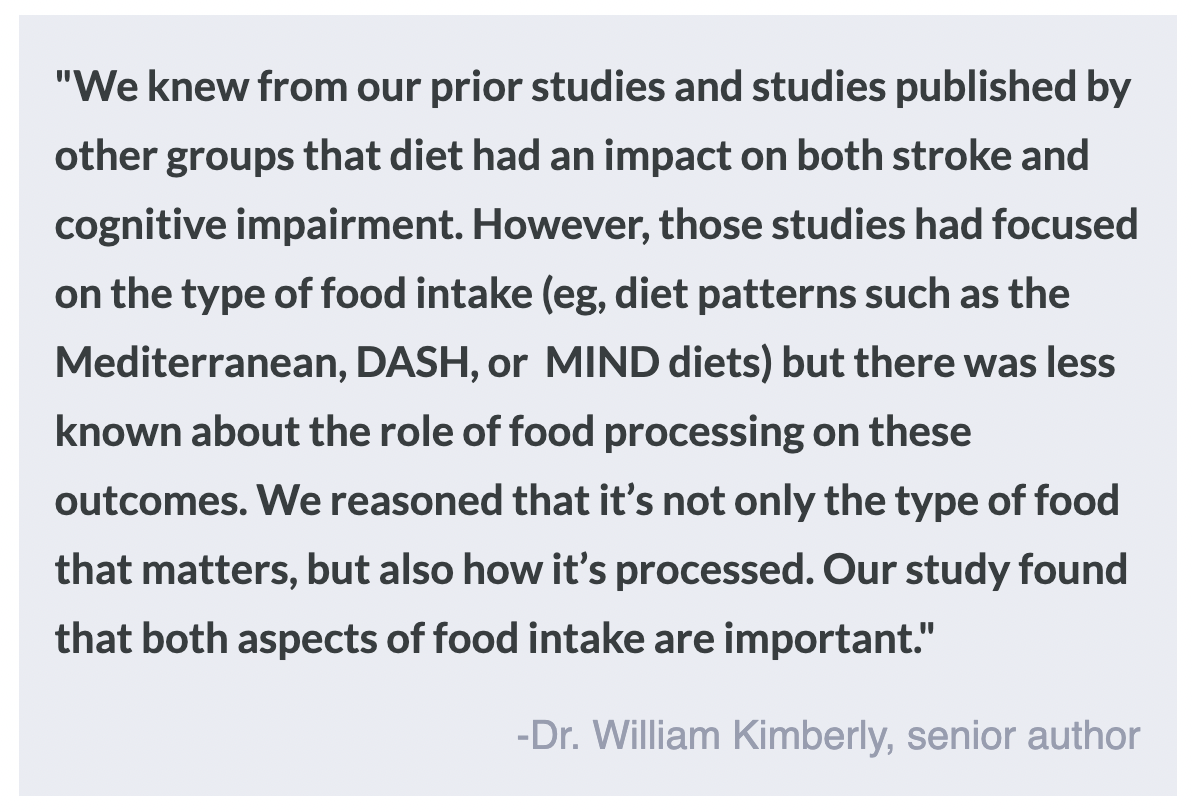
Diet is known to affect the brain - for example, following a more Mediterranean diet is associated with a reduced risk of stroke and lower cognitive decline. Recent research also indicates that eating more ultra-processed foods (e.g. carbonated drinks, flavoured yogurt, instant foods, packaged bread, chicken nuggets, etc.) is associated with a higher risk of stroke and faster cognitive decline. This week in Neurology, Bhave and colleagues published a study that adds to the growing literature on diet and brain health outcomes, investigating the role of food processing compared to following specific diets such as the Mediterranean diet. 

How did they do it?

The authors followed a cohort of over 30,000 non-Hispanic Black and White adults aged 45 and above in the United States, who entered the study between 2003 and 2007. After enrolling in the study, participants were assessed for clinical information, including a history of stroke or cognitive impairments, and demographic and lifestyle information. During the baseline assessment, participants answered a questionnaire about their food intake, which was then analyzed in two ways: 1) The food and drink items were categorized into four groups based on the level of processing, and daily intake for each category (in grams) was divided by total intake to get a proportion. 2) the questionnaires were scored based on how much they adhered to three healthy dietary patterns – Mediterranean, Dietary Approaches to Stop Hypertension (DASH), and Mediterranean-DASH Intervention for Neurodegenerative Delay (MIND).

After this baseline assessment, participants were followed up routinely to assess whether they had experienced a stroke, and to assess their cognitive performance in standardized tests. The authors then built statistical models to investigate the associations between food and drink intake and the incidence of stroke and cognitive impairment. 

What did they find?

The study found that eating more ultra-processed foods was associated with an increased risk of stroke, particularly in Black participants. On the other hand, eating more unprocessed/minimally processed foods or more strongly following a healthy diet was associated with a decreased risk of stroke. The results were similar when the authors looked at cognitive impairment – more ultra-processed foods were associated with greater cognitive decline, while less processed foods and healthier diets were associated with less decline.

The authors also asked a follow-up question: does the level of food processing matter if participants are following a healthier dietary pattern such as DASH or MIND? The answer was yes: even when participants adhered to a healthy diet, eating more ultra-processed foods was associated with some negative brain health outcomes, and eating more unprocessed foods was associated with better outcomes. This finding suggests the level of processing in the diet alone, is important for brain health, independently of other dietary patterns.

What's the impact?

This study highlights the important role that food processing plays in brain health. As always, it is important to note that these findings do not necessarily mean that eating more processed foods directly causes stroke and cognitive impairment (i.e. correlation is not causation). However, this study contributes to a growing literature that suggests a healthy diet including unprocessed food is important in maintaining brain health.