Connectivity of the Amygdala Predicts Risk Tolerance

What's the science?

Risk can be thought of as uncertainty — when there is some information about the possible outcome of a situation. Different individuals have different tolerance for risk when making decisions. We know that certain brain regions are generally involved in risk perception from studies looking at brain activation during risk (e.g. medial prefrontal cortex, anterior insula, anterior cingulate cortex, amygdala), however, we don’t know which brain regions and which inherent properties of these brain regions affect individual risk tolerance. This week in Neuron, Jung and colleagues use a data-driven approach to determine which brain regions and functional properties of these regions predict individual risk tolerance.

How did they do it?

Anatomical MRI, resting-state MRI (brain activity at rest) and Diffusion Tensor Imaging (structural connectivity) data from 108 healthy adults were acquired. Participants also performed a well-validated risk task to assess their risk tolerance. This task involves making binary decisions over several trials, choosing between a certain monetary reward and a larger uncertain (i.e. riskier) reward. They first analyzed the resting-state MRI data to compute individual functional connectivity throughout the brain (synchrony between brain regions at rest) to determine important regions that show a large amount of synchrony with other brain regions (i.e. highly central brain regions). In an exploratory, data-driven approach, they then assessed whether the strength of the functional connectivity in any these regions throughout the brain predicted individual risk tolerance.

What did they find?

The strength of functional connectivity in the amygdala showed the strongest correlation with risk tolerance of any brain region. Based on this finding, the authors focused on the amygdala for the remainder of their analyses. They tested which specific functional connections of the amygdala were important for risk tolerance. They used the amygdala as a seed region and found that the medial prefrontal cortex showed the strongest functional connections. There was a positive correlation between risk tolerance and functional connectivity between the amygdala and the medial prefrontal cortex; greater risk tolerance was associated with stronger functional connections. They then assessed whether the structural connectivity (white matter tracts) between the amygdala and the medial prefrontal cortex was associated with risk tolerance, and found that there was a negative correlation between structural connectivity and risk tolerance;  stronger white matter tract connectivity was associated with lower risk tolerance (significant for the right amygdala, and trending for the left amygdala). They also found that more gray matter volume in the amygdala was associated with a higher risk tolerance. In a regression analysis, they found that functional connectivity, gray matter volume and tract strength (only on the right) were all predictors of individual risk tolerance.

Amygdala functional connectivity and risk tolerance

What's the impact?

This is the first study to show that the inherent properties of the amygdala and its’ connections are associated with individual risk tolerance. This study suggests that an individual’s brain structure and function, which can be thought of as their “brain signature” can be used to predict individual behavior. Localizing brain regions involved in risk tolerance is important for understanding why some individuals engage in risk-taking behavior.

kable.png

W.H. Jung et al., Amygdala Functional and Structural Connectivity Predicts Individual Risk Tolerance. Neuron (2018). Access the original scientific publication here.

Functional Connections in the Brain are Stronger in Females Resilient to Depression

What's the science?

One third of females will be diagnosed with depression (major depressive disorder) during their adolescence. Resilience refers to the ability to adapt well in response to stress and bounce back from challenging life experiences. Currently, we don’t know the brain mechanisms that underlie resilience in adolescents who are at risk for depression. This week in JAMA Psychiatry, Fischer and colleagues test whether brain functional connectivity can be a biomarker for resilience in adolescent females at risk for depression (i.e. depression runs in their family).

How did they do it?

65 adolescent females were recruited: 25 low risk control participants who did not develop depression (control), 20 whose parents had a history of depression and developed depression themselves (i.e. converted) and 20 whose parents had a history of major depressive disorder but did not develop depression (i.e. resilient). The brains of all participants were scanned several times using  resting-state fMRI (which measures brain function at rest) over several years. They compared functional connectivity (synchronous brain activity) between resilient and converted females and between resilient and control females. They assessed the functional connectivity profiles of three brain regions known to be involved in depression: the amygdala (emotion), the anterior insula (attention/cognition) and the dorsolateral prefrontal cortex (planning). They measured the relationship between functional brain connections and life events.

What did they find?

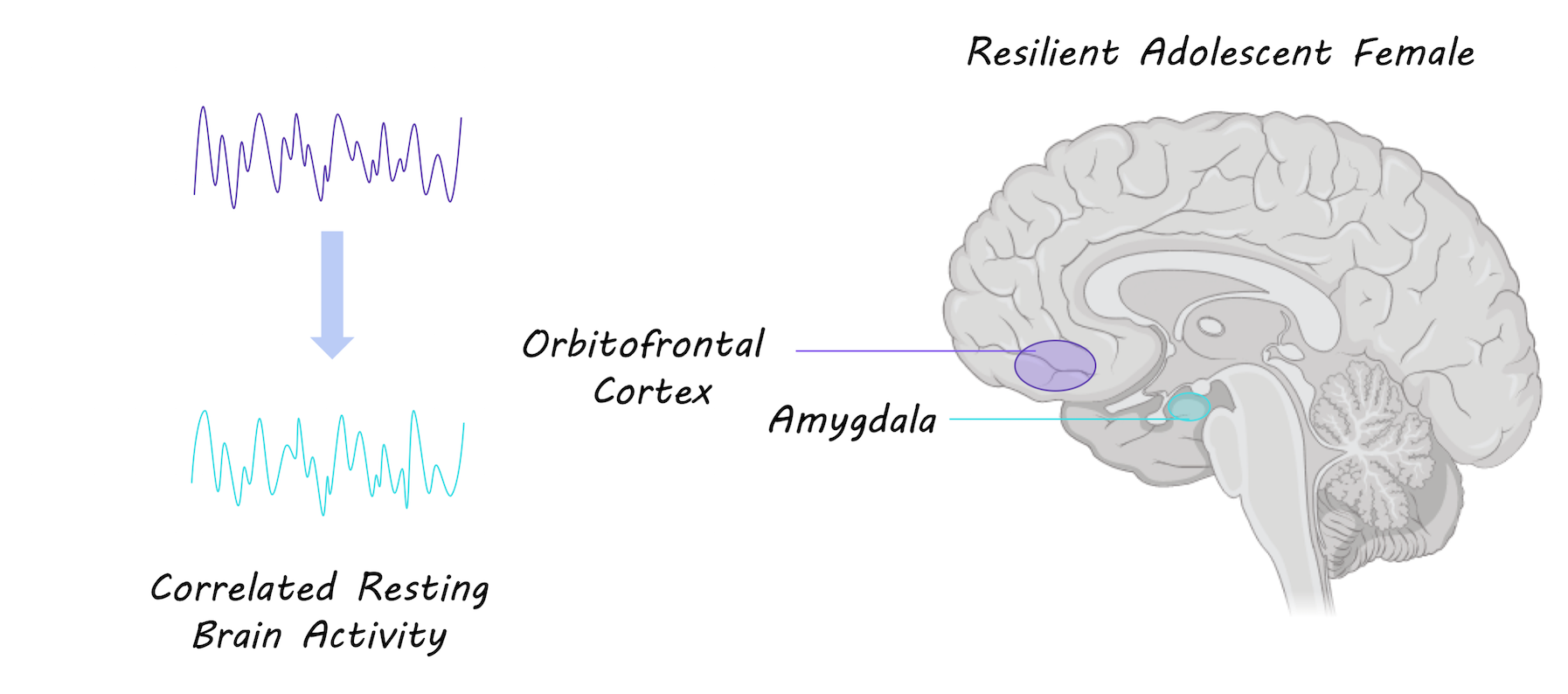
Females who were resilient to depression showed stronger functional connections in the brain between the amygdala (involved in fear and emotion) and the orbitofrontal cortex (involved in impulse control and modulating emotions). A stronger connection between these regions was associated with more positive life events. Resilient individuals also showed stronger connections between the dorsolateral prefrontal cortex (involved in planning and executive function) and the frontotemporal cortex (involved in cognitive control). Both resilient and converted groups had stronger functional connectivity within the salience network (a network of regions involved in attention and cognition) compared to the control group

Functional brain connectivity between orbitofrontal cortex and amygdala

What's the impact?

This is the first study to show that functional connections in the brain can be markers for resilience to depression in adolescent females at high risk for depression. Stronger functional connections could represent adaptation in the brain in response to positive life experience. It is crucial to understand how adolescents can develop resilience to depression in order to better prevent and treat depression.

inkquote4.png

A. Fischer et al., Neural Markers of Resilience in Adolescent Females at Familial Risk for Major Depressive Disorder. JAMA Psychiatry (2018). Access the original scientific publication here.

Your Brain Reacting to Social Injustice

What's the science?

How do we perceive injustice? Many neuroimaging studies have looked at how we perceive the violation of social norms by analyzing brain activity while participants play a computer game. For example, participants might have the option to punish one player who is acting unfairly (e.g. stealing) towards another. Further, different hormones, like oxytocin, influence our social behaviour, suggesting they can play a role in our perception of injustice. This week in The Journal of Neuroscience, Stallen and colleagues performed a new set of experiments using brain imaging to analyze the perception of injustice.

How did they do it?

First, oxytocin was administered to half of the participants. Next, all participants underwent an fMRI brain scan, while playing three computer games: 1) Participants played against an opponent, called a ‘taker’. The taker had the opportunity to steal up to 100 chips away from the participant, and the participant could then punish the taker by giving up up to 100 of their own chips. For each chip they gave up 3 would be taken from the taker, (injustice happening to them) 2) Participants received 200 chips and observed a taker stealing chips from another player, and could then punish the taker (using up to 100 chips, 3 taken from the taker for each chip given up), (observing social injustice, punishing as a third party) and 3) Participants observed a taker stealing chips from another player, and could compensate the disadvantaged player using up to 100 chips (the disadvantaged player was given 3 chips per chip given up) (observing social injustice, compensating as a third party). Participants knew they would receive real monetary compensation after the games according to their performance, and all games were anonymous.

Perception of injustice computer game

What did they find?

Participants who received oxytocin were more likely to dole out small punishments, frequently, to a taker who took chips from the participant or another player, versus participants who did not receive oxytocin. When the authors compared trials in which a participant doled out punishment to the taker versus compensating the disadvantaged player, there was greater activity in the ventral striatum -- a brain region involved in processing rewards. The decision to administer punishment was associated with activity in the anterior insula -- a brain region involved in “gut feelings” and decision making involving risk. Activity in the amygdala, a brain region associated with affective arousal, was correlated with the severity of punishment administered but only in experiment #2, when participants observed a taker behaving unfairly towards someone else.

What's the impact?

This is the first study to assess the perception of social justice in situations where an individual is experiencing injustice firsthand compared to observing injustice as a third party. This study suggests two distinct brain mechanisms might be at play during these unjust situations.

A word of caution: Different brain regions are activated in many different situations. Just because a brain region is known to be activated during reward, for example, does not necessarily mean that brain region will always be active during reward processing.

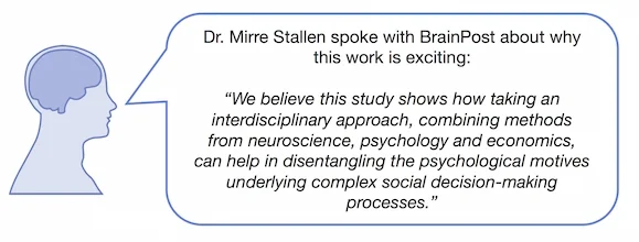
Stallen_comment_new_small.jpg

M. Stallen et al., Neurobiological Mechanisms of Responding to Injustice. Journal of Neuroscience. (2018). Access the original scientific publication here.