Connectivity of the Amygdala Predicts Risk Tolerance

What's the science?

Risk can be thought of as uncertainty — when there is some information about the possible outcome of a situation. Different individuals have different tolerance for risk when making decisions. We know that certain brain regions are generally involved in risk perception from studies looking at brain activation during risk (e.g. medial prefrontal cortex, anterior insula, anterior cingulate cortex, amygdala), however, we don’t know which brain regions and which inherent properties of these brain regions affect individual risk tolerance. This week in Neuron, Jung and colleagues use a data-driven approach to determine which brain regions and functional properties of these regions predict individual risk tolerance.

How did they do it?

Anatomical MRI, resting-state MRI (brain activity at rest) and Diffusion Tensor Imaging (structural connectivity) data from 108 healthy adults were acquired. Participants also performed a well-validated risk task to assess their risk tolerance. This task involves making binary decisions over several trials, choosing between a certain monetary reward and a larger uncertain (i.e. riskier) reward. They first analyzed the resting-state MRI data to compute individual functional connectivity throughout the brain (synchrony between brain regions at rest) to determine important regions that show a large amount of synchrony with other brain regions (i.e. highly central brain regions). In an exploratory, data-driven approach, they then assessed whether the strength of the functional connectivity in any these regions throughout the brain predicted individual risk tolerance.

What did they find?

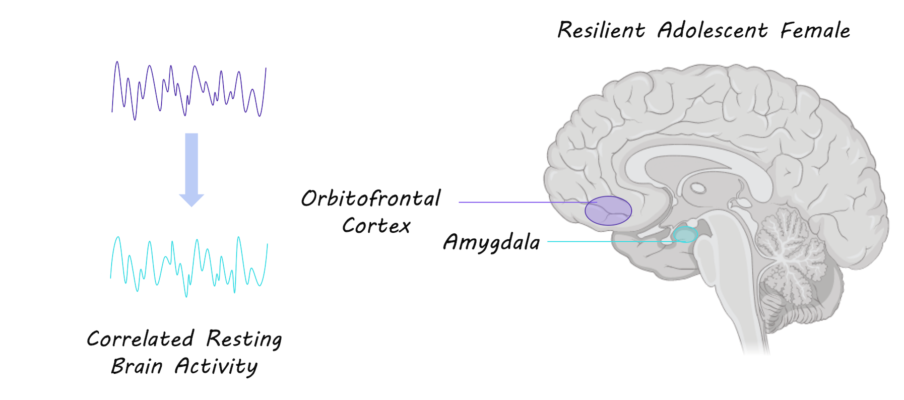
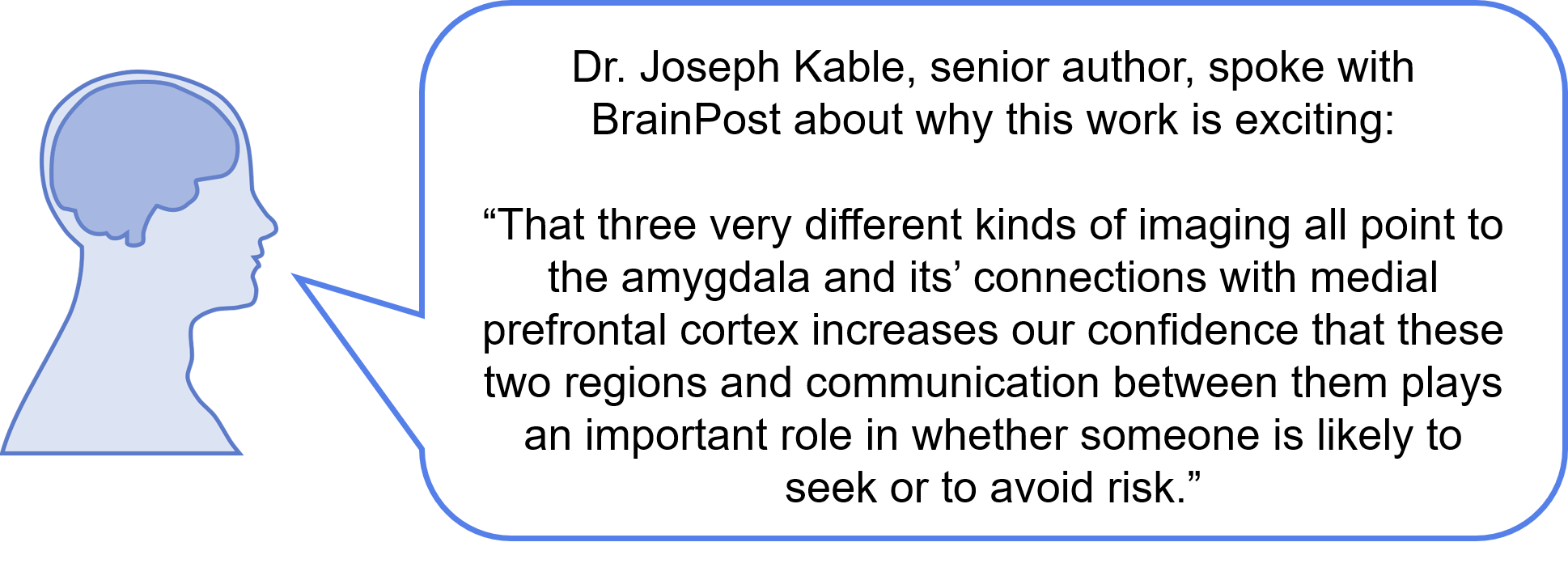
The strength of functional connectivity in the amygdala showed the strongest correlation with risk tolerance of any brain region. Based on this finding, the authors focused on the amygdala for the remainder of their analyses. They tested which specific functional connections of the amygdala were important for risk tolerance. They used the amygdala as a seed region and found that the medial prefrontal cortex showed the strongest functional connections. There was a positive correlation between risk tolerance and functional connectivity between the amygdala and the medial prefrontal cortex; greater risk tolerance was associated with stronger functional connections. They then assessed whether the structural connectivity (white matter tracts) between the amygdala and the medial prefrontal cortex was associated with risk tolerance, and found that there was a negative correlation between structural connectivity and risk tolerance;  stronger white matter tract connectivity was associated with lower risk tolerance (significant for the right amygdala, and trending for the left amygdala). They also found that more gray matter volume in the amygdala was associated with a higher risk tolerance. In a regression analysis, they found that functional connectivity, gray matter volume and tract strength (only on the right) were all predictors of individual risk tolerance.

Amygdala functional connectivity and risk tolerance

What's the impact?

This is the first study to show that the inherent properties of the amygdala and its’ connections are associated with individual risk tolerance. This study suggests that an individual’s brain structure and function, which can be thought of as their “brain signature” can be used to predict individual behavior. Localizing brain regions involved in risk tolerance is important for understanding why some individuals engage in risk-taking behavior.

kable.png

W.H. Jung et al., Amygdala Functional and Structural Connectivity Predicts Individual Risk Tolerance. Neuron (2018). Access the original scientific publication here.

Functional Connections in the Brain are Stronger in Females Resilient to Depression

What's the science?

One third of females will be diagnosed with depression (major depressive disorder) during their adolescence. Resilience refers to the ability to adapt well in response to stress and bounce back from challenging life experiences. Currently, we don’t know the brain mechanisms that underlie resilience in adolescents who are at risk for depression. This week in JAMA Psychiatry, Fischer and colleagues test whether brain functional connectivity can be a biomarker for resilience in adolescent females at risk for depression (i.e. depression runs in their family).

How did they do it?

65 adolescent females were recruited: 25 low risk control participants who did not develop depression (control), 20 whose parents had a history of depression and developed depression themselves (i.e. converted) and 20 whose parents had a history of major depressive disorder but did not develop depression (i.e. resilient). The brains of all participants were scanned several times using  resting-state fMRI (which measures brain function at rest) over several years. They compared functional connectivity (synchronous brain activity) between resilient and converted females and between resilient and control females. They assessed the functional connectivity profiles of three brain regions known to be involved in depression: the amygdala (emotion), the anterior insula (attention/cognition) and the dorsolateral prefrontal cortex (planning). They measured the relationship between functional brain connections and life events.

What did they find?

Females who were resilient to depression showed stronger functional connections in the brain between the amygdala (involved in fear and emotion) and the orbitofrontal cortex (involved in impulse control and modulating emotions). A stronger connection between these regions was associated with more positive life events. Resilient individuals also showed stronger connections between the dorsolateral prefrontal cortex (involved in planning and executive function) and the frontotemporal cortex (involved in cognitive control). Both resilient and converted groups had stronger functional connectivity within the salience network (a network of regions involved in attention and cognition) compared to the control group

Functional brain connectivity between orbitofrontal cortex and amygdala

What's the impact?

This is the first study to show that functional connections in the brain can be markers for resilience to depression in adolescent females at high risk for depression. Stronger functional connections could represent adaptation in the brain in response to positive life experience. It is crucial to understand how adolescents can develop resilience to depression in order to better prevent and treat depression.

inkquote4.png

A. Fischer et al., Neural Markers of Resilience in Adolescent Females at Familial Risk for Major Depressive Disorder. JAMA Psychiatry (2018). Access the original scientific publication here.

Resting Brain Activity Predicts Who Responds to Cognitive Behavioral Therapy for OCD

What's the science?

Obsessive Compulsive Disorder (OCD) affects 1-2% of the population and can affect quality of life. Cognitive behavioral therapy (CBT) is a method of treatment that has been shown to be effective in some individuals, but not all. Currently, there is no method to predict who will benefit from CBT. Recently, functional MRI of individuals at rest has emerged as a promising tool for predicting treatment outcomes. This week in PNAS, Reggente and colleagues test whether resting brain activity patterns can predict treatment response.

How did they do it?

Adults with a diagnosis of OCD underwent resting state functional MRI scans before and after 4 weeks of daily CBT. They analyzed the resting state fMRI scans using a multivariate approach and machine learning to detect whether patterns of resting state activity before treatment could predict individual OCD symptom severity scores after treatment. Resting brain activity was extracted from 196 brain regions and patterns of activity in all regions were correlated with one another. Multivariate analyses have the ability to capture multiple patterns of brain activity, and may be better than univariate approaches for predicting individualized responses to treatment. OCD symptom severity was also assessed before and after the 4 weeks of treatment.

What did they find?

OCD symptom severity scores improved after treatment in almost all participants. The authors found that pre-treatment resting state patterns in two brain networks -the default mode network and the visual network - strongly predicted individual variability in OCD symptom severity score. The default mode network (active while an individual is at rest) accounted for 67% of the variation in post-treatment symptom severity scores, while the visual network accounted for 51%. The activity in these networks better predicted post-treatment severity scores than the severity of OCD before treatment.

Brain by cronodon.com, Image by BrainPost

Brain by cronodon.com, Image by BrainPost

What's the impact?

Knowing who will respond to treatment is important as CBT is time consuming and expensive. This is the first study to report resting state network patterns as a reliable predictor of individual response to CBT treatment for obsessive compulsive disorder. Individual resting state patterns could reflect the plasticity or adaptability of brain networks to treatment. This study brings us one step closer to using individualized treatment plans for complex disorders.

Reggente_author_week5_small.jpg

Reach out to study author Dr. Nicco Reggente on Twitter @mobiuscydonia

N. Reggente et al., Multivariate resting-state functional connectivity predicts response to cognitive behavioral therapy in obsessive–compulsive disorder. PNAS. (2018). Access the original scientific publication here.